Tubo-ovarian Abscess: Pearls & Pitfalls
- Aug 12th, 2015
- Dina Al-Joburi
- categories:
Tubo-ovarian Abscess: Pearls & Pitfalls
Author: Dina Al-Joburi, DO
(EM Resident Physician, Drexel University College of Medicine)
Edited by: Alex Koyfman, MD (@EMHighAK, EM Attending Physician, UT Southwestern Medical Center / Parkland Memorial Hospital) and Stephen Alerhand, MD (@SAlerhand)
Case #1
12 year-old girl presents with five days of right lower quadrant abdominal pain, anorexia, nausea, and vomiting. Pain is constant and non-radiating. Patient is not pregnant and LMP was two weeks prior and regular. Past surgical history is significant for appendectomy two years prior. On physical examination, the patient is febrile to 102.4 F, abdominal exam reveals right lower quadrant tenderness and labs are significant for WBC of 13,000.
Outcome: patient found to have right TOA in the OR, right adnexa was adherent to the sigmoid colon. Presumed that appendectomy predisposed the patient to delayed development of TOA8
Case #2
61 year-old G1P1 presents with three weeks of post-menopausal vaginal bleeding and three days of abdominal pain and dysuria with past medical history of vaginal prolapse. On physical exam, patient febrile to 101.2, abdominal exam pertinent for suprapubic tenderness and pelvic exam reveals an erythematous cervix, but no adnexal mass palpated. Labs were significant for a WBC of 24,000.
Outcome: left TOA secondary to adenocarcinoma of the endometrium 9
Case #3
35-year-old G0 presents with diffuse abdominal pain, bilious emesis, and constipation for one day. Patient has no pertinent past medical or surgical history. On physical, the patient is afebrile, but her abdomen is slightly distended and diffusely tender. All labs were within normal limits.
Outcome: SBO secondary to bowel entrapment in left-sided TOA3
Introduction
Tubo-ovarian abscess is an inflammatory mass involving the fallopian tube, ovary, or adjacent pelvic organs. When there is an agglutination of these structures, it is called a tubo-ovarian complex.1 While TOA was once more commonly associated with being life-threatening, advancements in antibiotics and surgical techniques have resulted in a near absent mortality.1 However, failure to recognize it can result in irreversible tubal and ovarian damage, chronic pelvic pain, adhesion formation, ectopic pregnancy, and abscess rupture.
All of the above patients, despite their diverse presentations ended up getting diagnosed with tubo-ovarian abscesses. These cases underscore the point that unfortunately not all patients present with the slam-dunk presentation of fever, pelvic pain, and pelvic mass in a female with a history of PID. Depending on the patient’s demographics and presentation, specific diagnostic and treatment considerations must be taken.
Pathogenesis
TOA can be classified as either primary or secondary. The most widely accepted cause of a primary TOA is thought to be a result of an ascending infection from the lower genital tract. This infection, thought to be secondary to either sexually transmitted organisms or endogenous flora, ascends to the fallopian tube and causes an inflammatory response. Pus accumulates causing pyosalpingitis that extends to the ovary. A TOA may also result from local spread of infection from the appendix, bowel, bladder, or in association with a pelvic malignancy, this process is labeled a secondary TOA.10
Risks
The risks for TOA can be separated into PID-related and non-PID-related. The PID-related risks include the following: multiple sexual partners, age between 15-25, history of prior PID, and having an IUD in place.1 However, not all TOAs are associated with PID. TOA may also arise as a post-operative complication, therefore look for history of pelvic/abdominal surgery and ovarian manipulation as seen with in vitro fertilization. Patients with chronic inflammatory process of the bowel e.g. Crohn’s disease and ulcerative colitis, and even diverticulitis can be at risk for a TOA.1
Presentation
Presentations can be separated into the non-ruptured vs. ruptured TOA. Not all women present in an acute fashion. In fact, an un-ruptured abscess is more difficult to diagnose because of its variable clinical presentation.
Typical symptoms seen with intact abscesses include: vaginal discharge, acute lower abdominal pain, fever, irregular vaginal bleeding, and urinary symptoms. However, some women present with seemingly unrelated symptoms (as seen in the cases above), such as change in bowel movements, upper abdominal pain and back pain.
Ruptured tubo-ovarian abscess spans a spectrum ranging from abscess leak with localized peritoneal reaction and mild systemic changes to rupture with generalized peritoneal sepsis and septic shock.11
Differential Diagnoses
While the differential list is long and can span across the reproductive system, gastrointestinal tract, and urinary tract, key differentials to keep in mind in the ED are ectopic pregnancy, PID, ovarian torsion, appendicitis, and incarcerated hernia.
Approach
Labs include CBC, +/- ESR and CRP, pregnancy test and cervical cultures of gonococcus and chlamydia.
As clinical presentation can vary greatly and patients may or may not be febrile with leukocytosis, diagnosis is best made with pelvic ultrasound.
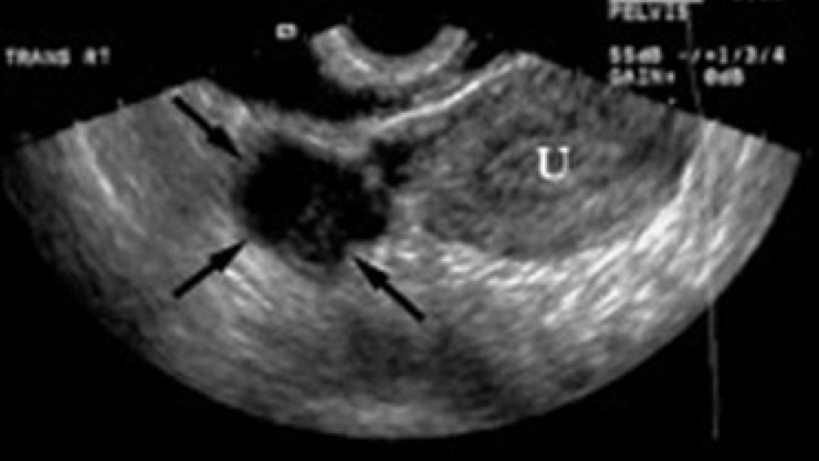
Ultrasound will show a complex, thick-walled, adnexal structure. However, more benign diagnoses can mimic TOAs, such as endometriomas and dermoid cysts. If radiologist does not read the ultrasound as a TOA, but there is a high index of suspicion, further evaluate with a CT. 10
Treatment and Disposition
Patients who are hemodynamically stable, premenopausal, and abscess <9cm in diameter are good candidates for antibiotic therapy alone.2 Antibiotic coverage should target N. gonorrhoeae, C. trachomatis, and anaerobes.
Surgical intervention should be strongly considered in patients with suspected TOA rupture, sepsis presumed to be secondary to the TOA and post-menopausal women with TOA.
All patients should be admitted to monitor response to antibiotics, as failure to respond to treatment within 48-72 hrs is an indication for drainage or surgical intervention.2
Note that, special care should be taken in certain patient populations, i.e. the sexually inactive, pre-menarche and post-menopausal, to elucidate the cause of the TOA. As mentioned earlier, the post-menopausal patient should be evaluated for possible malignancy. 5-7
Summary
–Failure to recognize TOA can result in irreversible tubal and ovarian damage
-TOA can be seen in the pregnant, sexually inactive, and post-menopausal patient
-Be on alert for malignancy in the post-menopausal patient
-Think beyond PID as a sole risk factor: look for a history of infertility treatments and pelvic surgery
-Clinical presentations vary
-If ultrasound read doesn’t match the clinical scenario, get further imaging
-Admit all patients to monitor for response to antibiotics
References and Further reading
1) Beigi, Richard H., MD. “Epidemiology, Clinical Manifestations, and Diagnosis of Tuboovarian Abscess.” Epidemiology, Clinical Manifestations, and Diagnosis of Tuboovarian Abscess. UpToDate, n.d. Web. 15 June 2015.
2) Beigi, Richard H., MD. “Management and Complications of Tuboovarian Abscess.” Management and Complications of Tuboovarian Abscess. UpToDate, n.d. Web. 15 June 2015.
3)) G. Pines, Y. Klein, A. Ben Arie, S. Machlenkin, H. Kashtan: Small bowel obstruction due to Tubo Ovarian Abscess. IMAJ, 10 (2008), pp. 481–482
4) Hartmann KA, Lerand SJ, Jay MS. Tubo-Ovarian Abscess in Virginal Adolescents: Look for Underlying Etiology. J Pediatr Adolesc Gynecol 2007; 20: 127.
5)Khan NA, Maajeeni EH. Tubo-ovarian abscess in a postmenopausal woman with underlying ovarian carcinoma. Saudi Med J 2005; 26: 1010-1011.
6) Protopapas, Athanasios G., Emmanuel S. Diakomanolis, Spyros D. Milingos, Alexandros J. Rodolakis, Sophia N. Markaki, Georgios D. Vlachos, Dimitrios E. Papadopoulos, and Stylianos P. Michalas. “Tubo-ovarian Abscesses in Postmenopausal Women: Gynecological Malignancy until Proven Otherwise?” European Journal of Obstetrics & Gynecology and Reproductive Biology 114.2 (2004): 203-09. Web.
7) Metz Y, Nagler J. Diverticulitis presenting as a tubo-ovarian abscess with subsequent colon perforation. World Journal of Gastrointestinal Surgery. 2011;3(5):70-72. doi:10.4240/wjgs.v3.i5.70.
8) Sacks DA, Niswander KR, Easton TH: Tubo-ovarian abscess at puberty. West J Med 127:513-514, Dec 1977
9) S Kremer, R Kutcher, R Rosenblatt, F McGill, A Rozenblit, and M Goldman: Postmenopausal tubo-ovarian abscess: sonographic considerations and clinical significance. JUM November 1992 11:613-6
10) Velcani, Artur, Patrick Conklin, and Neil Specht. “Sonographic Features of Tubo-ovarian Abscess Mimicking an Endometrioma and Review of Cystic Adnexal Masses.” Radiology Case Journal of Radiology Case Reports 4.2 (2010):
11) Weekes LR: Ruptured tubo-ovarian abscess. J of National Medical Association 1975, 67(6):436–443.

TOA is a difficult diagnosis to make and I think the info here is well put together. One thing to note is that US does not perform nearly as well as we have been led to believe. A colleague and I published a review of this topic about 6 years ago (http://www.ncbi.nlm.nih.gov/pubmed/20466506). Sensitivity may be as low as the 50s% but likely is in the high 70% range. CT has performed better but isn’t 100% either. Starting with an US is reasonable but progression to a CT should be strongly considered if the US is negative and suspicion is high. A negative CT may still require expert consultation and a possible OR trip.