The Cleared Trauma Patient: what could we be missing?
- May 6th, 2016
- Brendan Milliner
- categories:
Author: Brendan Milliner, MD (Senior EM Resident, Icahn School of Medicine at Mount Sinai) // Edited by: Stephen Alerhand, MD (@SAlerhand) and Alex Koyfman, MD (@EMHighAK, EM Attending Physician, UTSW / Parkland Memorial Hospital)
It’s early on a Monday morning. The crowds haven’t started to arrive in your ED yet, and you’re chatting with your co-workers, finishing your coffee, feeling calm and relaxed about the day ahead. Suddenly the phone rings, and it’s your department chair.
“Listen, do you remember that 20 year-old guy you discharged the other day after he was hit by a car? Turns out he went to see an orthopedist and was diagnosed with a Lisfranc. He’s pretty upset that we missed it. I’d like you to come by my office after your shift.”
That coffee doesn’t taste very good anymore. You try to remember the details –minor accident, negative ED workup – you even x-rayed the foot and didn’t see any fracture. Sent him out on crutches and instructions for ‘contusion.’ How could you have seen this coming?
As ED docs, we see our share of trauma. Depending on your practice environment, this could be anything from crashing poly-trauma to minor scrapes and fractures. If your shop is anything like mine, most of your patients are the latter and eventually get discharged from the ED after you do your best to exclude dangerous or disabling injuries.
What can we do to prevent scenarios like the one above? Some number of patients will inevitably leave your ED with an undiagnosed injury, but by understanding the types of injuries that are most commonly missed, we can help to reduce the number that slip through the cracks. In today’s article, we’ll be discussing musculoskeletal and abdominal injuries, as well as the special cases of central cord syndrome and diaphragmatic injury
Bad to the Bone: MSK
Most trauma patients receive some kind of workup for MSK injuries, typically tailored to symptoms and deformities noted on exam. A portable chest and pelvis is standard ACLS, and head/c-spine CT is usually included for significant head or neck trauma. Most ED docs will x-ray painful extremities in trauma, but the majority of missed MSK injuries come from falsely negative x-rays.
Several specific injuries are known as ‘easy to miss.’ These include
-
Lisfranc fracture-dislocation:
A Lisfranc refers to disrupted alignment of metatarsals with the tarsal bones. It often occurs in conjunction with metatarsal fracture. It’s a scary fracture for ED docs, as an estimated 20% go unrecognized1, and without surgery it can lead to permanent disability. Here’s what to look for:
- Tenderness along Lisfranc joint line
- Plantar ecchymosis
- Disruption of 2nd or 4th On a normal x-ray, the 2nd metatarsal should line up exactly with the cuneiform bone, and the 4th should line up with the cuboid.

- Caption: Normal alignment of the 2nd metatarsal

- Caption: Normal alignment of the 3rd and 4th metatarsal
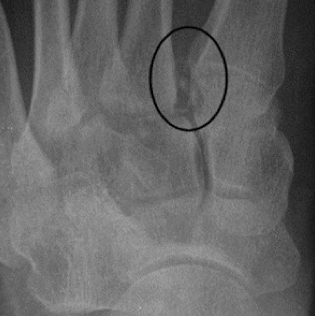
- ‘Fleck sign’. This is an avulsion fragment at the base of the metatarsal. Lateral displacement of the 2nd metatarsal can be seen
Images from emergencymedicinecases.com
If you see any of the above, get an Orthopedics consult. These injuries warrant acute orthopedic evaluation. If in doubt, weight-bearing films or (more likely) CT will help to clarify the diagnosis. Minor Lisfranc patients may be able to walk, so do not let that reassure you! For suspicious findings with unremarkable x-rays/CT, you can consider discharge with posterior splint, non-weight bearing instructions, and ortho referral.
-
FOOSH injuries
a. Scaphoid fracture
Even the med student knows to check for this one, and for good reason: scaphoid fracture is the most common carpal fracture (up to 70%) and can result in necrosis of the bone and disability. A recent review of the literature suggests that both x-rays and CT are insufficient to rule it out in the acute setting2 (MRI is better). So, here’s what to do:
- Check for snuffbox tenderness. This is one of those old-school teachings that plays out. The review mentioned above found that absence of snuffbox tenderness had an LR- of 0.15 for fracture, better than any other clinical sign.
- Don’t be reassured by X-rays or CT. See above.
- If you are concerned, splint the wrist and arrange for prompt Ortho follow-up.
b. Scapholunate dislocation
The awkward cousin of the scaphoid fracture, this injury involves ligamentous disruption between the scaphoid and lunate. It’s most common with a FOOSH. What to look for:
- Terry Thomas (or Madonna) sign: this refers to widening of >2 mm between the scaphoid and lunate on the AP x-ray.

- Image from radswiki.net
- Signet ring sign: Abnormal orientation of scaphoid. This is only seen in significant instability. You can see this in the above image; the scaphoid appears tilted compared to normal.
- If concerned, again: splint and send to Ortho
c. DRUJ (distal radio-ulnar joint dislocation)
Another FOOSH injury! Friends don’t let friends FOOSH. Be suspicious when there is pain over distal radius or ulna or deformity, either with or without associated fracture. Look for:
- Crepitus over distal ulna with movement of the wrist
- Prominence of ulnar styloid missing
- Widening of the joint or displacement of the ulna relative to the radius on x-ray. Most of the time, the ulna goes dorsally. You can see this well on a lateral x-ray (below) if it is a good film.

- Caption: There is volar dislocation of the distal radio-ulnar articulation. No associated fracture identified. Image from radiopedia.org.
- Big dislocations can be reduced in the ED. Supinate and put pressure over the ulnar head. Make sure to rule out a fracture first.
- Splint and send to Ortho. You guessed it!
-
C-spine injury
The debate about the best way to clear a c-spine has been raging since EM was in its infancy, and we’ll just touch on it briefly here. In my shop, patients are cleared with the NEXUS criteria if possible, and assessed with cervical CT if not.
Data regarding patients with continuing midline neck pain after a negative CT is mixed, and it’s probably safest to send these patients out with a soft collar and prompt spine follow-up. That said, a recent review in the neurosurgery literature including over 1,000 patients found no UNSTABLE ligamentous injuries initially not recognized that were found later with MRI. 3
Yo’ Stomach: Blunt abdominal injuries
Moving on to the next category of injuries: the abdomen. Abdominal trauma comes in two flavors: blunt and penetrating. The management of penetrating trauma to the abdomen is relatively straightforward, and your trauma team is unlikely to send home a patient with a knife or gunshot wound to the belly that requires an ex-lap.
Blunt trauma can be another story. What do you do with the construction worker that falls 10 feet onto concrete but appears stable without significant abdominal pain? Does he need a CT, admission to the Observation unit, or can you send him back to work?
As every in-service test-taker knows, the most commonly injured organs in blunt abdominal trauma are the spleen and liver, with hollow viscus or bladder injuries also possible. Your first step is a FAST, but this will only be positive in cases of significant intra-peritoneal hemorrhage, and there are no clear criteria as to which patients require further testing or Observation.
A recent retrospective study from Denver Health including 1169 patients4 sheds some light on this question. As per their protocol, Denver placed all trauma activations not immediately admitted into an Obs unit for eight hours with plan to repeat the abdominal exam and hematocrit in stable patients prior to discharge. Of these patients, only 3% were admitted to the hospital for suspected injury; the most common reason for admission was persistently abnormal vital signs. Only two patients were found to have a significant abdominal injury.
A second trial in 2012 out of UC Davis prospectively evaluated 2,734 patients with normal CT scans after blunt abdominal trauma5 (performed with IV contrast only). Of these, eight patients (0.3%) had a significant abdominal injury identified later. The majority of these patients either had abnormal abdominal exams or were altered.
So what does this tell us? First of all, the rate of intra-abdominal injury missed on initial eval seems to be very low, at least at these institutions. This assumes that your trauma assessment includes the standard physical exam, FAST, hematocrit, and urinalysis. In patients without a severe mechanism who do not have concerning findings or abnormal vitals, you are probably okay to send them home. If there’s something that worries you, CT or observation may pick up additional injuries.
Breathe, breathe in the air: Diaphragmatic injuries
Diaphragmatic injuries are one of the classic missed diagnoses in trauma patients. Caused either by penetrating trauma (~ 2/3 of cases) or severe blunt trauma (1/3), its presentation can be insidious. Some studies estimate the rate of missed injuries to be up to 66%.
- Look for: herniation of abdominal organs on x-ray or CT. X-ray has extremely poor sensitivity without large herniation, and even CT is not sensitive; reports range between 54-73%6.
- Missed diagnoses can result in bowel ischemia, so consider this in someone with recent severe trauma and abdominal pain/elevated lactate. The diaphragm does not heal well without repair.
- Detailed examination of the diaphragm intra-op is the best diagnostic tool
- Keep a high degree of suspicion. This is a tough one.
This feeling in my spine: Central cord syndrome
Traumatic central cord syndrome seems to be under-recognized among EM physicians and can lead to significant morbidity. Current theories suggest that acute hyperextension of the neck causes pinching and edema of the cervical cord, particularly in patients with significant DJD7. Unfortunately, this describes many elderly patients, a prime demographic for head trauma in the first place.

Illustration of the pathophysiology of central cord syndrome. Note the “pincer” effect on the central cord by anterior and posterior compression. Image from emedicine.medscape.com.
- Look for: weakness greater in the upper extremities, and less commonly urinary retention or sensory disturbance.
- All patients with neck trauma – particularly the elderly – should get a strength exam prior to discharge.
- Treatment for this condition is controversial and depends on severity, but can include acute spinal decompression7, so get Neurosurgery involved if you detect weakness.
That wraps up today’s discussion of occult injuries in cleared trauma patients. In admitted trauma patients, many surgery teams do a ‘tertiary survey’ to identify any missed injuries after the life-threatening trauma is treated. We can do our own version of that in the ED: take a minute before patting your patient on the back and handing him his papers to reassess and run through these injuries in your mind. It’ll help to keep your patients healthy (and avoid those calls from your chair/legal team).
References / Further Reading
- Perron AD, Brady WJ, Keats TE. Orthopedic pitfalls in the ED: Lisfranc fracture-dislocation. Am J Emerg Med. 2001;19(1):71-75.
- Carpenter CR, Pines JM, Schuur JD, Muir M, Calfee RP, Raja AS. Adult scaphoid fracture. Acad Emerg Med. 2014;21(2):101-121.
- Chew BG, Swartz C, Quigley MR, Altman DT, Daffner RH, Wilberger JE. Cervical spine clearance in the traumatically injured patient: is multidetector CT scanning sufficient alone? Clinical article. J Neurosurg Spine. 2013;19(5):576-581.
- Kendall JL, Kestler AM, Whitaker KT, Adkisson MM, Haukoos JS. Blunt abdominal trauma patients are at very low risk for intra-abdominal injury after emergency department observation. West J Emerg Med. 2011;12(4):496-504.
- Holmes JF, McGahan JP, Wisner DH. Rate of intra-abdominal injury after a normal abdominal computed tomographic scan in adults with blunt trauma. Am J Emerg Med. 2012;30(4):574-579.
- Kuo IM, Liao CH, Hsin MC, et al. Blunt diaphragmatic rupture – a rare but challenging entity in thoracoabdominal trauma. Am J Emerg Med. 2012;30(6):919-924.
- Molliqaj G, Payer M, Schaller K, Tessitore E. Acute traumatic central cord syndrome: a comprehensive review. Neurochirurgie. 2014;60(1-2):5-11.

One thought on “The Cleared Trauma Patient: what could we be missing?”