Authors: Kendrick Lim, DO (EM Resident Physician, Carolinas Medical Center – Charlotte, NC); Bryant Allen, MD (Associate Professor of Emergency Medicine, Carolinas Medical Center – Charlotte, NC) // Reviewed by: Sophia Görgens, MD (EM Physician, Yale University, CT); Cassandra Mackey, MD (Assistant Professor of Emergency Medicine, UMass Chan Medical School); Alex Koyfman, MD (@EMHighAK); Brit Long, MD (@long_brit)
Welcome to EM@3AM, an emDOCs series designed to foster your working knowledge by providing an expedited review of clinical basics. We’ll keep it short, while you keep that EM brain sharp.
A 27-year-old male presents to the ED with multiple gunshot wounds to the lower abdomen.
Initial vital signs are BP of 130/80, HR of 110, RR 18, SpO2 of 97% on RA, T 98.5°F. On primary survey, he has a patent airway, bilateral breath sounds, 2+ radial and dorsalis pedal pulses. GCS 15. On secondary survey, patient has 1 penetrating injury to the left lower quadrant of the abdomen and 1 to the right proximal thigh. When patient’s back is examined, there is 1 penetrating injury to the right flank and 1 to the right buttocks. Notably, he has gross hematochezia.
E-FAST is negative. Bedside abdominal and pelvic x-ray show no foreign bodies. CT imaging notable for a wound tract adjacent to the patient’s rectum with extraluminal free air.
Question: What is the diagnosis?
Answer: Rectal Injury
Background:
- Higher incidence in the military setting when compared to civilian setting.1
- In the civilian setting, often seen with high mechanism motor vehicle collisions, gunshot wounds, and foreign bodies.1
- Historically, managed with 4 Ds1
- Debridement
- Drainage
- Diversion
- Distal irrigation
- Over the last several decades, there has been rethinking of traditional dogma and movement toward primary repair.1
Anatomy:
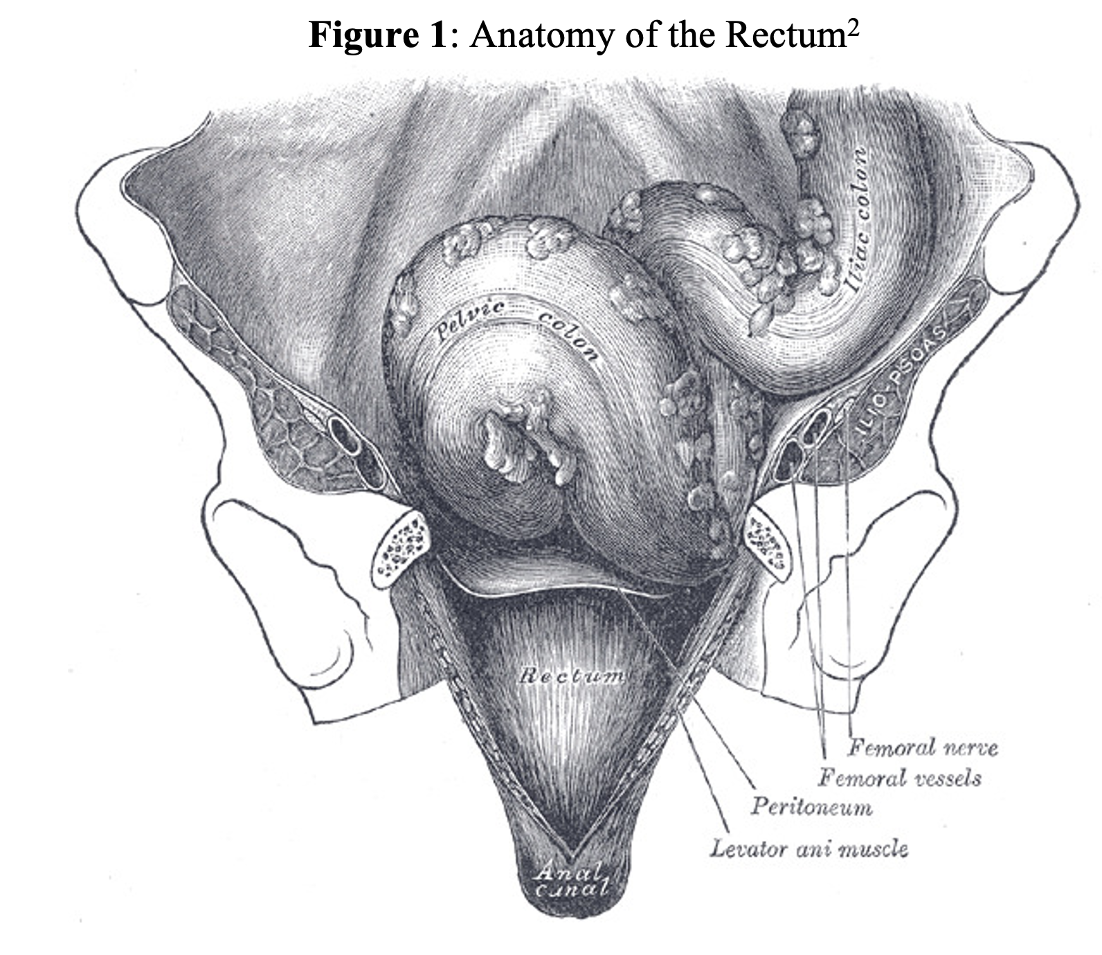
- The rectum is the most distal portion of the large intestine, bound by the sigmoid colon proximally and anal canal distally.2
- The rectum is approximately 12 to 15cm long.2
- It has an intraperitoneal portion, which includes the upper two-thirds anteriorly and the upper one third laterally. The lower third of the rectum is extraperitoneal.3
Epidemiology:
- Traumatic injuries to colon, rectum, and anus have primarily been studied and reported in the military setting.1
- Furthermore, most literature consists of prospective trials or retrospective case studies, limiting the exact incidence of these injuries.1
- During Operation Iraqi Freedom, colorectal injuries were identified in 175 (5.1%) of 3,442 injured service members.4
- Among 133 colorectal injuries sustained during Operation Iraqi Freedom, penetrating injury was the predominant mechanism of injury followed by blast (23%) and blunt trauma (5%).5
- In contrast, a retrospective civilian cohort of 227,972 patients presenting with blunt trauma identified colorectal injuries in only 798 (0.3%) patients.6
- Often devoid of high-energy/blast injuries seen in combat setting.6
- A systematic review of civilian colorectal trauma literature (1999-2016) found gunshot wounds to be the most common mechanism of injury (46.9%).7
- Most patients were male (>85%), and injuries were often associated with other pelvic fractures.7
Clinical Presentation:
- Generally, in the traumatic setting
- Blunt trauma with concern for pelvic fractures
- Penetrating injuries with trans-pelvic trajectory
- Foreign body insertion
- Pelvic instability, blood at the urethral meatus, soft-tissue defect of the perineum, or penetrating injury near the pelvis should heighten suspicion for rectal injury.
- Other symptoms include hematochezia, anorectal pain, abdominal pain.
Evaluation:
- Standard Adult Trauma Life Support Protocol
- Airway, breathing, circulation
- E-FAST ultrasound exam
- Digital Rectal Examination (DRE)
- In the ATLS setting, focus on sphincter tone, presence of blood, foreign objects, or bony protrusion — especially in those patients with pelvic fractures.
- Recent evidence has challenged the DRE as futile given its lack of sensitivity.8
- For spinal cord injury, 37% sensitive and 96% specific
- For bowel injury, 5.7% sensitive and 98.9% specific
- For rectal injury, 33.3% sensitive and 99.8% specific
- For pelvic fracture, 0% sensitive and 99.8% specific
- For urethral disruption, 20% sensitive and 99% specific
- Furthermore, DRE findings have been shown to alter management in fewer than 4% of patients.9
- Finally, there is a high risk of a negative and traumatic experience for patients, physical harm to clinicians performing the examination as well as risk for litigation, and transmission of infectious disease.9
- Overall, the DRE should not be routinely performed, but the rectal examination may have a higher probability of influencing management for those with penetrating injuries in proximity to the lower gastrointestinal tract, questionable spinal cord damage, and severe pelvic fractures with potential urethral disruption or open fractures in continuity with the rectal vault.10
- In these settings, concerning findings include a defect in the rectal wall, gross blood, decreased anal sphincter tone, bony fragments, or a high-riding prostate.11
- Proctoscopy
- Proctoscopy often does not detect a distinct injury but demonstrates less conclusive findings such as intraluminal blood, which may often prompt laparoscopy to evaluate for intra-abdominal rectal injury.11
- In a study of 77 patients, proctoscopy was 78% sensitive for rectal injuries with a 58% sensitivity for intraperitoneal injuries and 88% sensitivity for extraperitoneal injuries.12
- Computed Tomography (CT)
- There is sparse evidence regarding sensitivity and specificity of CT for rectal injuries.
- In a small retrospective study of 10 patients injured in combat, CT did not miss any rectal injuries. Notably, there was a 20% false-positive rate.13
- The use of rectal contrast is institution dependent but may not adequately evaluate the distal rectum due to occlusion by the device’s balloon.11
- Ultimately, there is an inadequate level of evidence to support or refute use of intraluminal contrast.11
Treatment:
- Trauma and/or general surgery consultation should be obtained early for all patients with a traumatic rectal injury, with anticipated plan for hospital admission.
- Other concurrent injuries are often present.
- Prophylactic antibiotics
- Broad-spectrum antibiotics targeting both aerobes and anaerobes preoperatively and 24 hours postoperatively.14
- The Eastern Association for the Surgery of Trauma (EAST) guidelines do not specify a preferred antibiotic regimen.15
- As per surgical team (often based on intra- vs extra-peritoneal involvement):1
- Primary repair
- Resection and anastomosis
- Exteriorizing the injured segment into an ostomy (historically first line management)
Prognosis:
- Historically, high morbidity and mortality rates (60-75% during World War I).
- With advances in trauma and surgical management, mortality has improved to 3-10% with complication rates of 18%.5,16
Pearls:
- Maintain a high suspicion for rectal injuries in the setting of trans-pelvic penetrating injuries and high mechanism blunt trauma to the pelvis.
- Digital rectal examination is highly unlikely to change management, even in the presence of a rectal injury.
- Involve trauma and surgical teams early if concern for rectal injury.

A 39-year-old man presents with rectal pain and bleeding after inserting part of a lamp into his rectum. The lamp is removed in the ED. He is hemodynamically stable, but after removal, he develops a small amount of rectal bleeding. What is the best next step in the care of this patient?
A) CT angiography of the abdomen and pelvis
B) Discharge with outpatient surgical follow-up
C) Flexible sigmoidoscopy
D) Repeat digital rectal examination in 4 hours
Answer: C
Flexible sigmoidoscopy allows direct visualization of the rectal mucosa and can identify lacerations, ulcerations, ischemia, and perforationfollowing removal of a rectal foreign body. Rectal foreign bodies can produce injury through direct mechanical trauma, pressure necrosis, or manipulation during attempted extraction. Rectal bleeding following removal raises concern for mucosal injury and warrants further evaluation even when the patient is hemodynamically stable and does not have peritoneal signs.
In a hemodynamically stable patient without evidence of perforation or peritonitis, transanal extraction is generally the initial approach. Adequate analgesia and procedural sedation facilitate relaxation of the anal sphincter. The patient can be placed in the lithotomy position, and gentle manual extraction may be attempted if the object can be safely grasped. Digital manipulation around the object may help break a vacuum seal and allow air to pass proximally.
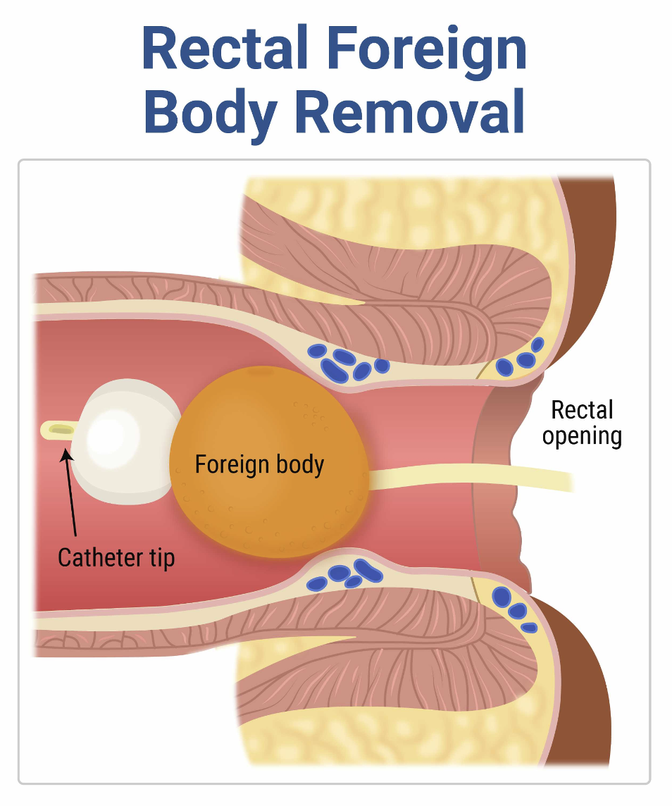
Various instruments can assist with extraction, including ring forceps, obstetric forceps, snares, and Foley catheters. A Foley catheter may be carefully passed beyond a smooth foreign body, the balloon inflated, and gentle traction applied to help bring the object distally. Endoscopic retrieval with a snare or other retrieval device can be considered for objects that are more proximal or cannot be removed manually. Objects made of glass require particular caution because forceful manipulation or grasping can shatter the object and produce severe rectal injury.
Repeated or forceful bedside attempts should be avoided. If transanal extraction fails, the foreign body is located high in the rectum or sigmoid colon, or safe extraction cannot otherwise be accomplished, examination under anesthesia with operative removal may be necessary. Laparoscopic or open abdominal surgery may be required for proximal objects that cannot be advanced distally or when significant bowel injury is identified.
The severity of injury determines subsequent care. Superficial mucosal injuries can generally be managed conservatively with observation and appropriate follow-up. In contrast, evidence of a full-thickness injury or perforation requires prompt surgical consultation, broad-spectrum antibiotics with gram-negative and anaerobic coverage, and operative management when indicated. Findings concerning for perforation include severe or worsening abdominal pain, peritoneal signs, fever, hemodynamic instability, and extraluminal or intraperitoneal free air.
Imaging also plays an important role when perforation is suspected. CT of the abdomen and pelvis can identify free air, free fluid, abscess formation, bowel wall abnormalities, and other complications. However, in this stable patient with no reported peritoneal findings after the foreign body has already been removed, direct endoscopic evaluation is best for identifying occult rectal mucosal injury.
CT angiography of the abdomen and pelvis (A) is primarily used to evaluate suspected active vascular bleeding and is not the preferred imaging modality for a stable patient with a small amount of rectal bleeding following foreign body removal. This patient has no hemodynamic instability or evidence of substantial ongoing hemorrhage. If perforation were suspected, a conventional CT of the abdomen and pelvis would be more appropriate than CT angiography.
Discharge with outpatient surgical follow-up (B) is inappropriate because rectal bleeding after foreign body removal raises concern for traumatic mucosal injury. A successful extraction does not exclude laceration, ischemia, or perforation. This patient should undergo additional evaluation before discharge to identify any clinically significant injury and ensure that no complications are developing. Flexible sigmoidoscopy provides direct assessment of the rectal mucosa.
Digital rectal examination (D) may identify gross blood, sphincter abnormalities, or a retained low-lying foreign body, but it cannot reliably exclude more proximal or subtle rectal injuries. Repeating the examination after several hours provides little additional diagnostic information and unnecessarily delays definitive care. Because this patient has rectal bleeding after foreign body removal, direct visualization of the mucosa with flexible sigmoidoscopy is more appropriate.
Further Reading/FOAMed:
StatPearls – https://www.ncbi.nlm.nih.gov/books/NBK551636/
References:
- Steele SR, Maykel JA, Johnson EK. Traumatic injury of the colon and rectum: the evidence vs dogma. Dis Colon Rectum. 2011;54(9):1184-1201. doi:10.1007/DCR.0b013e3182188a60
- Wang YHW, Wiseman J. Anatomy, Abdomen and Pelvis, Rectum. In:StatPearls. Treasure Island (FL): StatPearls Publishing; July 25, 2023.
- Mahan ME, Toy FK. Rectal Trauma. In:StatPearls. Treasure Island (FL): StatPearls Publishing; July 4, 2023.
- Steele SR, Wolcott KE, Mullenix PS, et al. Colon and rectal injuries during Operation Iraqi Freedom: are there any changing trends in management or outcome?Dis Colon Rectum. 2007;50(6):870-877. doi:10.1007/s10350-007-0235-4
- Cho SD, Kiraly LN, Flaherty SF, Herzig DO, Lu KC, Schreiber MA. Management of colonic injuries in the combat theater.Dis Colon Rectum. 2010;53(5):728-734. doi:10.1007/DCR.0b013e3181d326fd
- Williams MD, Watts D, Fakhry S. Colon injury after blunt abdominal trauma: results of the EAST Multi-Institutional Hollow Viscus Injury Study.J Trauma. 2003;55(5):906-912. doi:10.1097/01.TA.0000093243.01377.9B
- Ahern DP, Kelly ME, Courtney D, Rausa E, Winter DC. The management of penetrating rectal and anal trauma: A systematic review.Injury. 2017;48(6):1133-1138. doi:10.1016/j.injury.2017.03.002
- Shlamovitz GZ, Mower WR, Bergman J, et al. Poor test characteristics for the digital rectal examination in trauma patients. Ann Emerg Med. 2007;50(1):25-33.e1. doi:10.1016/j.annemergmed.2007.01.022
- Esposito TJ, Ingraham A, Luchette FA, et al. Reasons to omit digital rectal exam in trauma patients: no fingers, no rectum, no useful additional information. J Trauma. 2005;59(6):1314-1319. doi:10.1097/01.ta.0000198375.83830.62
- Porter JM, Ursic CM. Digital rectal examination for trauma: does every patient need one? Am Surg. 2001;67(5):438-441.
- Clemens MS, Peace KM, Yi F. Rectal Trauma: Evidence-Based Practices.Clin Colon Rectal Surg. 2018;31(1):17-23. doi:10.1055/s-0037-1602182
- Hargraves MB, Magnotti LJ, Fischer PE, et al. Injury location dictates utility of digital rectal examination and rigid sigmoidoscopy in the evaluation of penetrating rectal trauma.Am Surg. 2009;75(11):1069-1072. doi:10.1177/000313480907501108
- Johnson EK, Judge T, Lundy J, Meyermann M. Diagnostic pelvic computed tomography in the rectal-injured combat casualty.Mil Med. 2008;173(3):293-299. doi:10.7205/milmed.173.3.293
- Luchette FA, Borzotta AP, Croce MA, et al. Practice management guidelines for prophylactic antibiotic use in penetrating abdominal trauma: the EAST Practice Management Guidelines Work Group. J Trauma. 2000;48(3):508-518. doi:10.1097/00005373-200003000-00024
- Goldberg SR, Anand RJ, Como JJ, et al. Prophylactic antibiotic use in penetrating abdominal trauma: an Eastern Association for the Surgery of Trauma practice management guideline.J Trauma Acute Care Surg. 2012;73(5 Suppl 4):S321-S325. doi:10.1097/TA.0b013e3182701902
- Glasgow SC, Steele SR, Duncan JE, Rasmussen TE. Epidemiology of modern battlefield colorectal trauma: a review of 977 coalition casualties.J Trauma Acute Care Surg. 2012;73(6 Suppl 5):S503-S508. doi:10.1097/TA.0b013e3182754759