Authors: Catherine Durbin, MD (Emergency Medicine Resident, University of Kentucky); Blake Davidson, MD (Emergency Medicine Assistant Professor and Assistant Program Director, University of Kentucky) // Reviewed by: Sophia Görgens, MD (EM Physician, Yale University, CT); Cassandra Mackey, MD (Assistant Professor of Emergency Medicine, UMass Chan Medical School); Brit Long, MD (@long_brit); Alex Koyfman, MD (@EMHighAK)
Welcome to EM@3AM, an emDOCs series designed to foster your working knowledge by providing an expedited review of clinical basics. We’ll keep it short, while you keep that EM brain sharp.
A 28-year-old female presents to the emergency department with complaints of fatigue, bruising, and vaginal bleeding. She reports a history of heavy menstrual cycles; however, this cycle has been significantly worse. Over the past week, she has experienced dizziness, generalized fatigue, and increased bruising. She felt warm earlier this morning but was not able to take her temperature. She denies any family history of bleeding disorders.
Initial vital signs include BP 110/70, HR 120, RR 14, SpO2 100% on RA, and T 100.5F. On exam, she has diffuse petechiae and ecchymoses. Labs reveal hemoglobin 6.5 g/dL, WBC 1.6 x 10⁹/L, and platelet count of 40 x 109/L.
What is the diagnosis?
Answer: Aplastic Anemia
Etiology:
- Definition
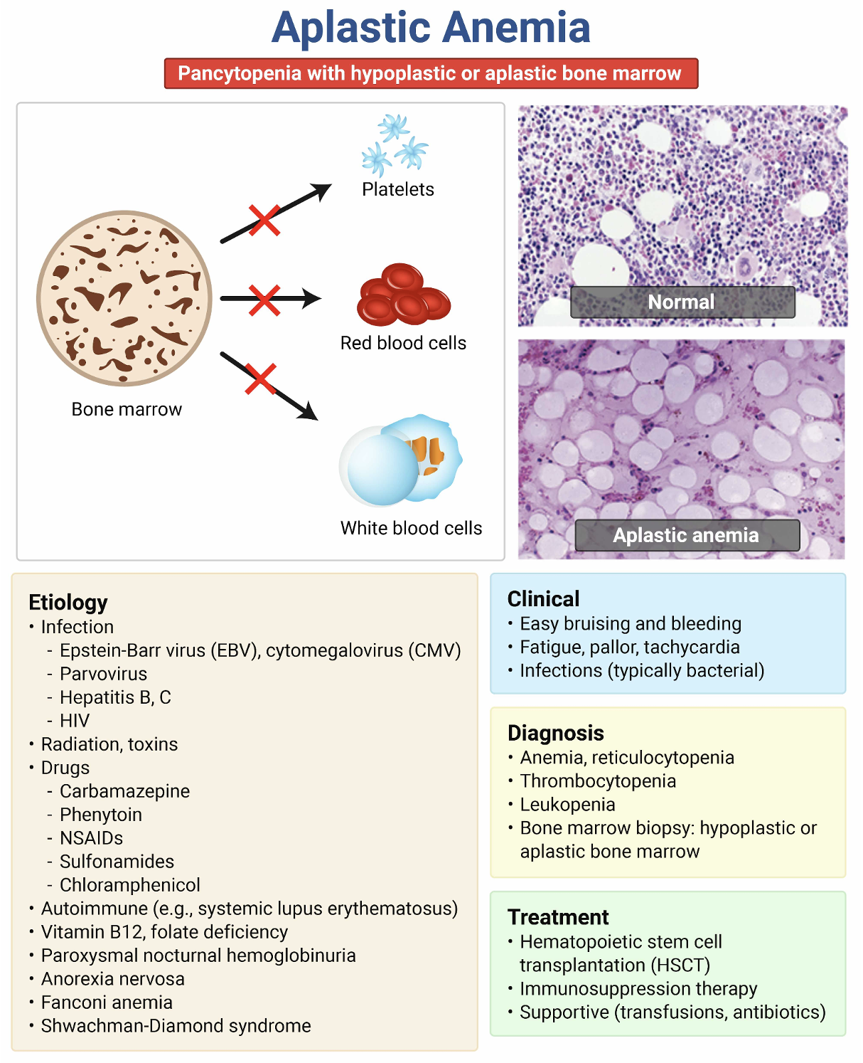
- Pancytopenia caused by a failure of the bone marrow to produce blood cells.1
- Pathophysiology of Aplastic Anemia1
- Chemical or physical damage (from toxins, drugs, or infections) causing replacement of bone marrow with fat
- Immune destruction of bone marrow via T cells
- Defects in genes essential for cell integrity and immune regulation.
- There are congenital and acquired etiologies.
| Congenital2 | Acquired |
| Fanconi Anemia | Drugs: Indomethacin, Diclofenac, Sulfonamides, Furosemide, Corticosteroids, Allopurinol, etc.3 |
| Congenital Keratosis | Infections: EBV, CMV, HIV, Varicella Zoster, HHV-6, and seronegative hepatitis4
|
| Congenital Pure Red Cell Aplasia | Toxins: Benzene, Pentachlorophenol, Lindane, Toluene, and organic solvents4
|
| Schwachman-Diamond Syndrome | Radiation |
| Autoimmune Disorders: Systemic Lupus Erythematous, Eosinophilic Fasciitis, Graft-Versus-Host-Disease4
|
Epidemiology:
- Incidence: 0.6-6.1 cases for every 1 million people4
- Equal ratio of affected males to females4
- Higher incidence in western Asia countries (Thailand, Taiwan)4
- Bi-modal distribution, peaking in the first 30 years of life and in those older than 604
Clinical Presentation:
- Can present over days, weeks or months5
- Symptoms5
- Exertional dyspnea, fatigue, easy bruising, epistaxis, heavy menstrual cycles, headache, fever, and gingival bleeding
- Physical Exam 5
- Pale complexion, petechiae, ecchymoses, and lack of lymphadenopathy and hepatosplenomegaly
Evaluation:
- Differential Diagnoses6
- Myelodysplastic syndromes, inherited bone marrow failure syndromes, large granular lymphocytosis, pure red cell aplasia, paroxysmal nocturnal hemoglobinuria, and myeloproliferative neoplasms
- ED evaluation:
- Complete Blood Count: normochromic, normocytic anemia with thrombocytopenia
- Hemoglobin: ↓, White Blood Cells: ↓, Platelets: ↓
- Peripheral Blood Smear7
- Platelets: ↓, Monocytic Percentage: ↓, Lymphocytic Percentage: ↑
- No blasts or malignant cells.
- Reticulocyte Count: ↓
- Comprehensive Metabolic Panel: typically normal, unless there are complications from the disease itself
- LDH: typically, normal
- Haptoglobin: typically, normal
- PT/INR: typically, normal
- APTT: typically, normal
- Type and Cross
- Complete Blood Count: normochromic, normocytic anemia with thrombocytopenia
- In the Hospital/Outpatient Setting
- Bone marrow biopsy: hypocellularity with little to no CD34+ cells8

-
- Cytogenetics
- Fluorescence In Situ Hybridization (FISH)
- Diagnosis
- Must have at least two of the following:10
- Hemoglobin Concentration < 10 g/dL
- Platelet Count < 50 x 109/L
- Neutrophil Count < 1.5 x 10⁹/L
- Must have at least two of the following:10
- Classification 11
- Severe
- Neutrophils < 0.5 x 10⁹/L, Platelets < 20 x 109/L, Reticulocytes < 20 x 109/L
- Very Severe
- Above criteria, but with neutrophils < 0.2 x 10⁹/L
- Non-severe
- Hypocellular bone marrow, but does not meet the above criteria
- Severe
Treatment:
- Non-severe cases or asymptomatic patients may not require treatment.
- Immunosuppression Therapy12
- Antithymocyte globulin: 40 mg/kg/day for four days
- Cyclosporine: 5 mg/kg/day
- Hematopoietic Cell Transplantation
- Transfusion5
- pRBCs: symptomatic anemia, hemoglobin < 7 g/dL
- Platelets: active bleeding, or platelets < 10 x 109/L
- At platelets < 10 x 109/L, there is increased risk of spontaneous intracranial hemorrhage.
- All transfusions need to be irradiated to prevent graft-versus-host-disease.
- Platelets: active bleeding, or platelets < 10 x 109/L
- pRBCs: symptomatic anemia, hemoglobin < 7 g/dL
- Neutropenic Fever13
- Neutrophils are important in combatting invasion of organisms. Therefore, their absence can lead to recurrent and severe infections.
- Initiate infectious work-up including blood cultures, chest x-ray, urinalysis, +/- CT imaging, +/- lumbar puncture
- The amount of evaluation will be patient dependent.
- First-line antibiotic therapy is typically cefepime or piperacillin-tazobactam.
- Antivirals, antifungals, and granulocyte colony-stimulating factor (G-CSF) can be considered on a case-to-case basis.
Complications:
- Bleeding, infections, transformation to lymphoproliferative disorders, and osteonecrosis of the hip4
Disposition:
- Most likely admit, unless they are classified as non-severe.
- A hematology consult is likely indicated in all cases.
Prognosis:
- Varies based on age, reticulocyte and lymphocyte counts, existence of anti-HLA antibodies, and complications of stem cell transplant.4
- Younger patients with severe aplastic anemia have survival rates around 80-90%.4
Pearls:
- A key diagnostic feature is the presence of hypocellular bone marrow with pancytopenia, not just anemia.
- Given neutropenia, patients are at increased risk for infections. Therefore, prophylaxis is recommended.
- Although the majority of cases are idiopathic, obtain a thorough drug and exposure history.
- Ensure that all transfused products are irradiated to reduce the risk of graft-versus-host disease.
- Have a low threshold to obtain CT in a thrombocytopenic patient with a headache.

A 44-year-old woman presents to the ED with fatigue and dyspnea. Physical examination shows cutaneous petechiae. Laboratory studies show pancytopenia. Reticulocyte count is 0.2%. The medical record shows a complete blood count within normal limits 6 months ago. Which of the following, if present, most likely contributed to this patient’s current condition?
A) Carbamazepine use
B) Glucose-6-phosphate dehydrogenase deficiency
C) Hepatic cirrhosis
D) Infection with Escherichia coli O157:H7
E) Iron deficiency
Answer: A
Aplastic anemia is a rare bone marrow failure syndrome caused by idiosyncratic immune-mediated destruction of hematopoietic stem cells. Roughly one-fourth of cases are drug induced, with carbamazepine, chloramphenicol, NSAIDs, gold salts, and antithyroid medications as the most common culprits. Other triggers include viral infections (hepatitis, parvovirus B19, Epstein-Barr virus), autoimmune disorders, and radiation exposure, though many cases are idiopathic. Patients present with features reflecting pancytopenia, including fatigue, dyspnea, bleeding or bruising (including petechiae), and fever or infections. The onset may be abrupt or insidious.
Definitive diagnosis will not be made in the ED, as it requires peripheral pancytopenia and hypocellular bone marrow, but suggestive laboratory findings should heighten suspicion during ED evaluation. Pancytopenia with reticulocytopenia is essential, as the latter reflects marrow failure rather than peripheral cellular destruction. ED workup should also include a peripheral smear, serum chemistries, coagulation studies, and type and screen to help exclude alternate diagnoses. Hematology involvement will likely also be necessary, but the urgency will vary depending on the severity of the presentation or on whether alternative diagnoses (especially thrombotic thrombocytopenic purpura) are suspected.
ED priorities include recognizing life-threatening cytopenias and initiating supportive care while arranging urgent hematology consultation when appropriate. Immediate management should include supportive transfusions (RBCs and platelets as needed), treatment and prevention of infections, and avoidance of additional marrow-suppressing medications. Definitive treatment will be implemented in conjunction with specialty evaluation and might consist of immunosuppression or hematopoietic stem cell transplantation. Major acute complications include life-threatening infections and bleeding, with severe neutropenia conferring the highest mortality risk.
Glucose-6-phosphate dehydrogenase (G6PD) deficiency (B) causes episodic hemolytic anemia triggered by oxidative stress (e.g., infections, fava beans, certain drugs), not pancytopenia. Patients with hemolysis due to G6PD deficiency would have elevated reticulocyte counts rather than the reticulocytopenia seen here, and only the erythrocyte line would be affected.
While cirrhosis (C) can cause cytopenias through hypersplenism and decreased thrombopoietin production, it typically causes mild to moderate reductions in blood counts, not the severe pancytopenia with reticulocytopenia characteristic of marrow failure.
E. coli O157:H7 (D) causes hemolytic uremic syndrome (HUS), characterized by microangiopathic hemolytic anemia, thrombocytopenia, and acute kidney injury. HUS presents with hemolysis (elevated reticulocytes, schistocytes, elevated lactate dehydrogenase, low haptoglobin) and kidney dysfunction, not the pancytopenia with reticulocytopenia and hypocellular marrow seen here.
Iron deficiency (E) causes isolated microcytic anemia, sometimes with reactive thrombocytosis, but not pancytopenia. Reticulocyte counts may be low-normal in iron deficiency, but white blood cell counts and, usually, platelet counts remain normal.
Further Reading
https://pedemmorsels.com/aplastic-anemia/
https://radiopaedia.org/articles/aplastic-anaemia?lang=us
References:
- Young N. S. (2018). Aplastic Anemia. The New England journal of medicine, 379(17), 1643–1656. https://doi.org/10.1056/NEJMra1413485
- Wang, L., & Liu, H. (2019). Pathogenesis of aplastic anemia. Hematology, 24(1), 559–566. https://doi.org/10.1080/16078454.2019.1642548
- Gale, R.P., Hinterberger, W., Young, N.S. et al. What causes aplastic anaemia? Leukemia 37, 1191–1193 (2023). https://doi.org/10.1038/s41375-023-01892-2
- Nimmana BK, Penney SW. Aplastic Anemia. [Updated 2025 Jul 7]. In: StatPearls [Internet]. Treasure Island (FL): StatPearls Publishing; 2026 Jan-. Available from: https://www.ncbi.nlm.nih.gov/books/NBK534212/
- Brodsky, R. A., & Jones, R. J. (2005). Aplastic anaemia. The Lancet, 365(9471), 1647–1656. https://doi.org/10.1016/s0140-6736(05)66515-4
- DeZern, A. E., & Sekeres, M. A. (2014). The challenging world of cytopenias: distinguishing myelodysplastic syndromes from other disorders of marrow failure. The oncologist, 19(7), 735–745. https://doi.org/10.1634/theoncologist.2014-0056
- Elghetany, M. T., Hudnall, S. D., & Gardner, F. H. (1997). Peripheral blood picture in primary hypocellular refractory anemia and idiopathic acquired aplastic anemia: an additional tool for differential diagnosis. Haematologica, 82(1), 21–24.
- Amy E. DeZern, Jane E. Churpek; Approach to the diagnosis of aplastic anemia. Blood Adv 2021; 5 (12): 2660–2671. doi: https://doi.org/10.1182/bloodadvances.2021004345
- Bakhshi, S. (2025, December 19). Aplastic Anemia Workup. Approach Considerations, Complete Blood Cell Count and Peripheral Smears, Peripheral Blood Testing. https://emedicine.medscape.com/article/198759-workup#c9
- Kulasekararaj A, Cavenagh J, Dokal I, Foukaneli T, Gandhi S, Garg M, et al. Guidelines for the diagnosis and management of adult aplastic anaemia: A British Society for Haematology Guideline. Br J Haematol. 2024;204(3):784–804. https://doi.org/10.1111/bjh.19236
- Frickhofen, N., Kaltwasser, J. P., Schrezenmeier, H., Raghavachar, A., Vogt, H. G., Herrmann, F., Freund, M., Meusers, P., Salama, A., Heimpel, H., & German Aplastic Anemia Study Group. (1991). Treatment of Aplastic Anemia with Antilymphocyte Globulin and Methylprednisolone with or without Cyclosporine. New England Journal of Medicine, 324(19), 1297–1304. https://doi.org/10.1056/nejm199105093241901
- Peffault de Latour, R., Kulasekararaj, A., Iacobelli, S., Terwel, S. R., Cook, R., Griffin, M., Halkes, C. J. M., Recher, C., Barraco, F., Forcade, E., Vallejo, J.-C., Drexler, B., Mear, J.-B., Smith, A. E., Angelucci, E., Raymakers, R. A. P., de Groot, M. R., Daguindau, E., Nur, E., … Risitano, A. M. (2022). Eltrombopag Added to Immunosuppression in Severe Aplastic Anemia. New England Journal of Medicine, 386(1), 11–23. https://doi.org/10.1056/nejmoa2109965
- Embaby, Mostafa M.a; Metwalley, Kotb A.a; Riad, Rania S.b,. Febrile neutropenia in nonmalignant conditions: tertiary center experience. Journal of Current Medical Research and Practice 7(4):p 287-291, Oct–Dec 2022. | DOI: 10.4103/jcmrp.jcmrp_30_22