Author: Rachel Ely, DO, MHA, EMT-P (EM Resident at SAUSHEC, USAF) // Edited by: Alex Koyfman, MD (@EMHighAK, EM Attending Physician, UTSW / Parkland Memorial Hospital) and Brit Long, MD (@long_brit)
Case
A 61-year-old male presents to your ED with one week of epigastric pain and vomiting. He tells you that he had a cholecystectomy earlier this year, and his current pain is very similar to what prompted his gallbladder removal. He also says that his girlfriend mentioned that his eyes seemed “yellow”, and he complains of whole-body pruritis. He has vomited multiple times at home, and he is having difficulty answering your questions because of persistent nausea. His vitals on presentation are BP 171/99, HR 70, RR 16, oral temp 98.5, SpO2 99%. On exam, you notice obvious scleral and sublingual icterus, as well as generalized jaundice of the skin. His abdomen is mildly distended, with tenderness in the right upper quadrant without guarding or rebound tenderness. What should you be considering? What are the keys to evaluation and management?
Introduction
Cholangitis is a life-threatening infection of the biliary tract. It was first described in 1877, and for many decades the mortality secondary to cholangitis approached 100%.1 Identification and treatment of cholangitis has significantly improved over the last century, however there is still progress to be made. Challenges include not only early recognition of biliary infection, but also identification of those patients requiring emergent versus urgent biliary decompression.2 With these advances in identification and management, mortality has been cut to approximately 10%.1
For another great case and background on cholangitis, please see http://www.emdocs.net/cholangitis-deadly-cause-of-right-upper-quadrant-abdominal-pain/
General Physiology & Risk Factors
Cholangitis is a result of biliary obstruction and bacterial growth in the bile.1 Bacteria, primarily E. coli, Klebsiella species, and Enterococcus species, are thought to ascend from the duodenum into the common hepatopancreatic duct in the setting of ductal obstruction.3 Intraductal pressures increase as a result of obstruction, leading to a disruption of the tight junctions in the hepatic cellular architecture and serving as a route for bacteria to enter the bloodstream.1,4 Characteristics which predispose patients to the development of cholangitis include primarily bile duct stones and manipulation of the biliary tree such as ERCP or surgery. Less common risk factors include orthotopic liver transplantation, primary sclerosing cholangitis, and AIDS-related cholangiopathy.1,4 Malignancy is a common cause of biliary obstruction; however, it is less likely to develop cholangitis primarily as a result of malignant obstruction rather than as a result of biliary manipulation to relieve a malignant obstruction.4 Patients with indwelling biliary stents, or who have recently had instrumentation of their biliary tree, are more likely to have infection with Enterococcus, Pseudomonas, MRSA, or VRE.5
Presentation
Charcot’s triad of fever, right upper quadrant pain, and jaundice yields a 25% sensitivity for the detection of ascending cholangitis.6 Reynold’s pentad, which adds hypotension and altered mental status to the classic triad, is present in only of 5-7% of cases.4 The most common presenting signs and symptoms are fever, which is observed in about 90% of patients, and abdominal pain, seen in approximately 80% of patients. Cholangitis should be suspected in patients with fever, altered mental status, occult sepsis, or an otherwise unexplained elevated bilirubin, especially in the setting of biliary stones or recent biliary instrumentation. Because cholangitis is nearly uniformly fatal without appropriate treatment, it must remain high on the differential in adult patients with unexplained fever.7,8
Diagnosis
While no specific laboratory study is in itself indicative of cholangitis, elevation of liver function tests can be suggestive. Specifically, elevation of both GGT and alkaline phosphatase is approximately 90% sensitive for acute cholangitis.8 While elevations of white blood cell count, ESR, and CRP are non-specific, in the right clinical context this is highly suggestive of overwhelming systemic disease.9 Up to 70% of patients with cholangitis will have positive blood cultures, and while the IDSA does not recommend routine blood culture collection in mild community-acquired intra-abdominal infection, it should certainly be considered in those with severe disease or recent instrumentation.4,10
CT is the imaging study of choice to visualize the biliary system in suspected cholangitis. While ultrasound is often the preferred modality to visualize the biliary system, CT can better characterize complications such as hepatic abscess and can identify external sources of obstruction such as malignancy.4,7,11 CT carries an 87% sensitivity in actually identifying the etiology of biliary obstruction.12 Findings such as intra- and extrahepatic ductal dilation, duct wall thickening, liver enhancement, and the presence of gallstones can be suggestive of cholangitis. The finding of papillitis, or inflammation at the ampulla of Vater, and the presence of an ampullary stone on CT can also be indicative of cholangitis.13
Diagnostic Criteria and Classification
Because elevated LFTs, leukocytosis, and imaging evidence of biliary dilation can be indicative of other disease processes (such as cholecystitis or choledocholithiasis), attempts have been made to develop a set of diagnostic criteria for cholangitis. The Tokyo Guidelines are the best-known criteria and offer a sensitivity of 91.8% and specificity of 77.7% in derivation studies, though external validation studies have yet to be published.14,15 Three diagnostic categories are used to apply the tool: indicators of systemic inflammation, evidence of cholestasis, and imaging evidence. One indicator of systemic inflammation plus either imaging evidence or cholestasis evidence should prompt strong suspicion of cholangitis. Criteria to indicate inflammation are fever or shaking chills, white blood cell count less than 4,000 or greater than 10,000, or C-reactive protein greater than 1. Cholestasis can be indicated by total bilirubin greater than or equal to 2 mg/dL, or alkaline phosphatase, GGT, AST, or ALT above 1.5 times the upper limit of normal. Finally, imaging is considered confirmatory if biliary dilation or other evidence of a likely inciting etiology of obstruction and subsequent cholangitis, such as biliary stent, stricture, or malignancy.14
Table 1 TG13 Diagnostic Criteria for Acute Cholangitis.14
| Category | Threshold |
| A. Systemic Inflammation | |
| Fever or Shaking Chills | Body Temperature >38°C |
| Laboratory evidence of inflammatory response | WBC <4,000 or >10,000
CRP >1 |
| B. Cholestasis | |
| Jaundice | T-Bili ³2 mg/dL |
| Abnormal LFT | Alk Phos >1.5 x upper limit normal
GGT >1.5x upper limit normal AST >1.5x upper limit normal ALT >1.5x upper limit normal |
| C. Imaging | |
| Biliary dilatation | |
| Evidence of etiology on imaging | |
| Diagnosis should be suspected if one item from A plus one item from B or C are present
Diagnosis is considered definite if one item from, A, B and C are present. |
|
Treatment
Early identification and appropriate management of cholangitis is imperative because of its impressive mortality risk if not aggressively treated. Often, these patients present with evidence of sepsis and should be treated accordingly with fluid resuscitation and antibiotics. However, ultimately the presence of biliary obstruction is the factor allowing cholangitis to occur, and this obstruction must be relieved. While convention teaches that earlier biliary decompression is better, there have been some attempts to stratify the severity of disease in order to prioritize the urgency of decompression, as discussed below.
Antibiotic selection
As discussed, the most common organisms isolated in ascending cholangitis are E. coli, Klebsiella species, and Enterococcus species, but because these patients have often had recent biliary instrumentation and likely other nosocomial exposures, initial antibiotic therapy should remain broad. A carbapenem, piperacillin-tazobactam, cefepime plus metronidazole, ampicillin/sulbactam, or ceftriaxone plus metronidazole are great options, with the addition of vancomycin in those with likely health-care associated biliary infection.16 The most recent Tokyo treatment guidelines, published in 2013, warn against the use of fluoroquinolones because of increasing regional E. coli resistance to these agents.10
The Tokyo Guidelines recommend a severity-based hierarchy of antibiotic selection. Criteria for grading disease can be found in the table below. Mild or Grade I disease might be managed with a cephalosporin or ertapenem, or a fluoroquinolone in regions where resistance patterns are favorable. Grade II disease options include piperacillin/tazobactam, a third or fourth generation cephalosporin, or ertapenem. Finally, antibiotic coverage for severe (Grade III) disease or health-care associated infections include piperacillin/tazobactam, third or fourth generation cephalosporin, imipenem/cilastatin or meropenem, aztreonam plus metronidazole, plus vancomycin in addition to any of these options.4,10
Table 2 TG13 Severity Criteria for Acute Cholangitis14
| Grade III: Severe
Dysfunction in at least one of following systems: |
Criteria |
| Cardiovascular | Hypotension requiring dopamine ³5 mcg/kg/min, or any dose of norepinephrine |
| Neurologic | |
| Respiratory | PaO2/FiO2 ratio <300 |
| Renal | Oliguria, or serum creatinine >2.0 mg/dl |
| Hepatic | INR > 1.5 |
| Hematologic | Platelet Count <100,000/mm3 |
| Grade II: Moderate
Any two of the following |
|
| Abnormal WBC count | >12,000/mm3 or <4,000/mm3 |
| High Fever | ³39°C |
| Advanced Age | ³75 years |
| Hyperbilirubinemia | Total bilirubin ³5 mg/dL |
| Hypoalbuminemia | <70% lower limit normal |
| Grade I: Mild | |
| Does not meet criteria of Severe or Moderate at diagnosis. | |
Biliary Decompression
In certain cases of mild infection, patients may respond well to medical management alone. However, this remains the exception rather than the rule, and identifying these patients remains a challenge that requires further study. The 2007 and 2013 Tokyo Guidelines suggest that their disease stratification, discussed above, may aid in the decision of performing immediate or delayed biliary decompression, or whether decompression is necessary at all in cases of mild disease.6,10 The TG13 authors make no hard and fast recommendations about severity-based management, other than emphasizing that those with Grade III disease require near-immediate biliary decompression.10
Disposition
In nearly all cases, patients with cholangitis will require admission, and many will require ICU care for continued resuscitation and vasopressor support in cases of septic shock. However, source control is important to changing the course of disease, and this will require relief of the biliary obstruction. The most common option is bile duct stenting or stone removal via endoscopic retrograde cholangiopancreatography (ERCP).2 In cases in which this is not possible, such as in malignant obstruction or extremely unstable patients, percutaneous drainage of the gallbladder may be a better temporizing measure.17 Open surgical biliary decompression carries a higher morbidity and mortality than ERCP, but may be required in certain unique situations.4 In most cases the first consult should be to gastroenterology, as ERCP is the typical first step in decompression, however it is important to recognize that inpatient care is likely to be multidisciplinary, potentially including general surgery, gastroenterology, interventional radiology, and medical or surgical intensivists.
Case Conclusion
Your patient is not febrile, and the white blood cell count is 10.6. Knowing this, you reflexively move cholangitis lower on your differential list. However, the direct bilirubin returns at 7.0, and an alkaline phosphatase of 894 supports an obstructive lesion. The lab seems to be having difficulty analyzing the lipase because of the elevated bilirubin. You start with an ultrasound to evaluate for retained or spontaneous common bile duct stone; however, this study is grossly normal with the exception of heterogeneity of the pancreatic head, thought to be incidental. Seeking an explanation for the patient’s pain and obvious jaundice, you order a CT of the abdomen and pelvis, and you are surprised when you see this image:
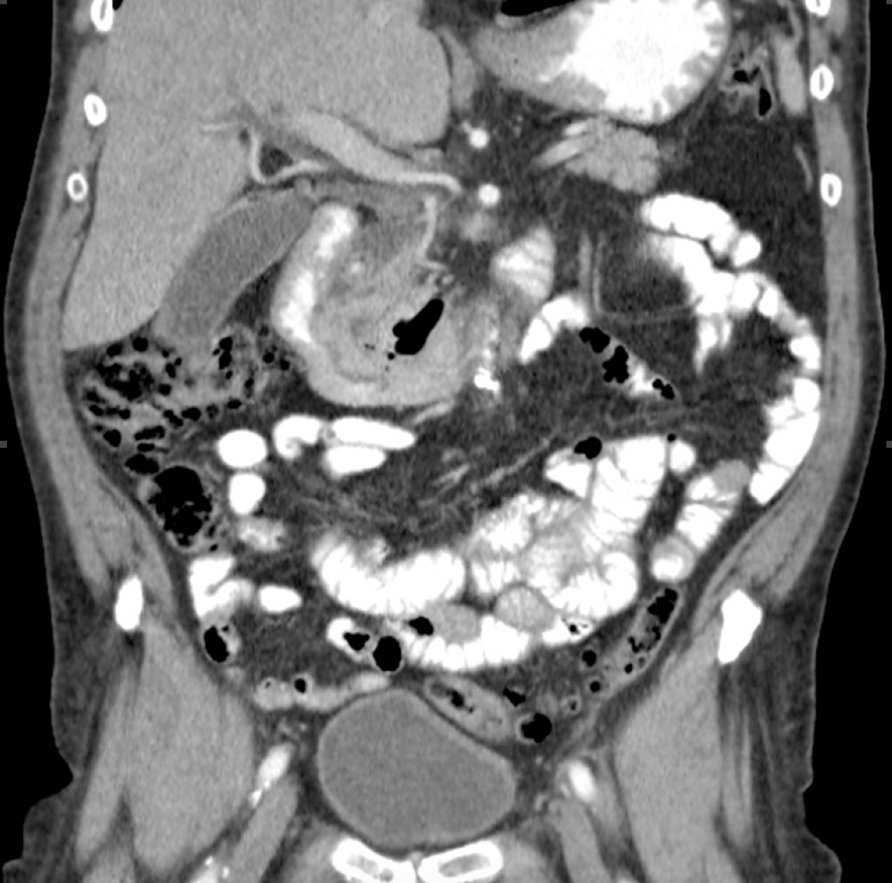
CT demonstrating choledocholithiasis and extra- and intra-hepatic ductal dilatation. Case courtesy of Dr. Roberto Schubert, Radiopaedia.org, rID/ 16704.
Your radiologic skills don’t typically allow you to pick out the common bile duct on CT, and the inflammation you see here clues you in that this patient may actually be harboring infection on top of his biliary obstruction. The lack of fever and leukocytosis initially threw you, but you soon realize that this patient meets the three diagnostic criteria for cholangitis per the Tokyo guidelines. You start piperacillin/tazobactam and metronidazole, give a fluid bolus, and call gastroenterology for consult and internal medicine for admission. When you follow up on the patient a few days later, you learn that the patient underwent ERCP with sphincterotomy, and stones and purulent fluid were drained from the common bile duct, confirming the diagnosis of cholangitis. His cholangitis and concurrent pancreatitis quickly resolved, and he is discharged on hospital day five.
Pearls & Takeaways
- Cholangitis can be subtle and only rarely presents with the classic triad of fever, jaundice, and right upper quadrant pain
- While ultrasound can be useful, CT is often required to support the diagnosis and can often identify the source of obstruction
- While early antibiotics are important, most patients require biliary decompression
- Consult gastroenterology early, and the patient may require ICU-level care
- The Tokyo Guidelines (TG13) may help support your working diagnosis in subtle cases, and can also help to stratify severity of disease. In the future, we may be able to use this severity stratification to guide urgency of biliary decompression.
References/Further Reading
1 Kimura Y, Takada T, Kawarada Y, Nimura Y, Hirata K, Sekimoto M, et al. Definitions, pathophysiology, and epidemiology of acute cholangitis and cholecystitis: Tokyo Guidelines. J Hepatobiliary Pancreat Surg 2007;14:15–26. doi:10.1007/s00534-006-1152-y.
2 Salek J, Livote E, Sideridis K, Bank S. Analysis of risk factors predictive of early mortality and urgent ERCP in acute cholangitis. J Clin Gastroenterol 2009;43:171–5. doi:10.1097/MCG.0b013e318157c62c.
3 Van den Hazel SJ, Speelman P, Tytgat GNJ, Dankert J, Van Leeuwen DJ. Role of antibiotics in the treatment and prevention of acute and recurrent cholangitis. Clin Infect Dis 1994;19:279–86. doi:10.1093/clinids/19.2.279.
4 Attasaranya S, Fogel EL, Lehman GA. Choledocholithiasis, Ascending Cholangitis, and Gallstone Pancreatitis. Med Clin North Am 2008;92:925–60. doi:10.1016/j.mcna.2008.03.001.
5 Tanaka A, Takada T, Kawarada Y, Nimura Y, Yoshida M, Miura F, et al. Antimicrobial therapy for acute cholangitis: Tokyo Guidelines. J Hepatobiliary Pancreat Surg 2007;14:59–67. doi:10.1007/s00534-006-1157-6.
6 Kiriyama S, Takada T, Strasberg SM, Solomkin JS, Mayumi T, Pitt HA, et al. New diagnostic criteria and severity assessment of acute cholangitis in revised Tokyo guidelines. J Hepatobiliary Pancreat Sci 2012;19:548–56. doi:10.1007/s00534-012-0537-3.
7 Block J, Lawrence LM FR. The Atlas of Emergency Radiology. In: Block J, Jordanov MI, Stack LB TR, editor. Atlas Emerg. Radiol., New York, NY: McGraw-Hill; 2013, p. Chapter 7.
8 Mosler P. Diagnosis and management of acute cholangitis. Curr Gastroenterol Rep 2011;13:166–72. doi:10.1007/s11894-010-0171-7.
9 Qin Y-S, Li Q-Y, Yang F-C, Zheng S-S. Risk factors and incidence of acute pyogenic cholangitis. Hepatobiliary Pancreat Dis Int 2012;11:650–4. doi:10.1016/S1499-3872(12)60240-9.
10 Gomi H, Solomkin JS, Takada T, Strasberg SM, Pitt HA, Yoshida M, et al. TG13 antimicrobial therapy for acute cholangitis and cholecystitis. J Hepatobiliary Pancreat Sci 2013;20:60–70. doi:10.1007/s00534-012-0572-0.
11 Schubert R. Choledocholithiasis | Radiology Case | Radiopaedia.org n.d. http://radiopaedia.org/cases/choledocholithiasis-4.
12 Balthazar EJ, Birnbaum BA, Naidich M. Acute cholangitis: CT evaluation. J Comput Assist Tomogr n.d.;17:283–9.
13 Lee NK, Kim S, Lee JW, Kim CW, Kim GH, Kang DH, et al. Discrimination of suppurative cholangitis from nonsuppurative cholangitis with computed tomography (CT). Eur J Radiol 2009;69:528–35. doi:10.1016/j.ejrad.2007.11.031.
14 Kiriyama S, Takada T, Strasberg SM, Solomkin JS, Mayumi T, Pitt HA, et al. TG13 guidelines for diagnosis and severity grading of acute cholangitis (with videos). J Hepatobiliary Pancreat Sci 2013;20:24–34. doi:10.1007/s00534-012-0561-3.
15 Yokoe M, Takada T, Mayumi T, Yoshida M, Hasegawa H, Norimizu S, et al. Accuracy of the Tokyo Guidelines for the diagnosis of acute cholangitis and cholecystitis taking into consideration the clinical practice pattern in Japan. J Hepatobiliary Pancreat Sci 2011;18:250–7. doi:10.1007/s00534-010-0338-5.
16 Solomkin, Joseph S. et al. Diagnosis and management of complicated intra-abdominal infection in adults and children: Guidelines by the Surgical Infection Society and the Infectious Diseases Society of America. Clin Infect Dis 2010;50:133–64. doi:10.1086/649554.
17 Lee CC, Chang IJ, Lai YC, Chen SY, Chen SC. Epidemiology and prognostic determinants of patients with bacteremic cholecystitis or cholangitis. Am J Gastroenterol 2007;102:563–9. doi:10.1111/j.1572-0241.2007.01095.x.


1 thought on “Cholangitis: Pearls & Pitfalls”
Pingback: emDOCs.net – Emergency Medicine EducationEM@3AM - Ascending Cholangitis - emDOCs.net - Emergency Medicine Education