Authors: Yenimar Ventura, MD; Muhammad Waseem, MD, MS (Lincoln Medical Center, Bronx New York) // Reviewed by: Tim Montrief, MD (@EMinMiami); Alex Koyfman, MD (@EMHighAK); Brit Long, MD (@long_brit)
Case
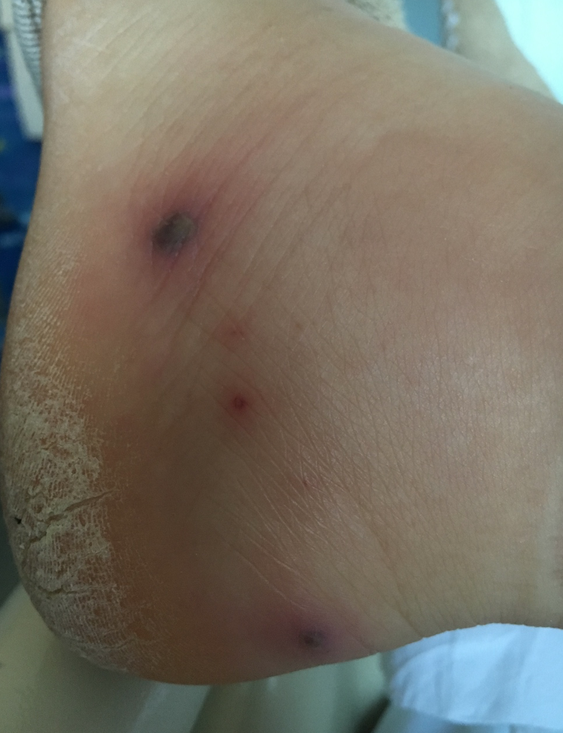
An 18-year-old girl presents with a 3-day history of fever, pruritic rash, and bilateral ankle pain and swelling associated with difficulty walking. On arrival, she has a fever of 101.4o F. A rash is present on her lower legs, feet, and the right palm. (Figure 1). Her left ankle has diffuse swelling, warmth, redness, tenderness, and a limited range of motion. What is on your differential diagnosis and what is the best management for this patient?

Disseminated Gonococcal Infection (DGI)
Disseminated Gonococcal Infection (DGI) is the most severe form of infection by Neisseria gonorrhoeae. It is a manifestation of acute systemic gonococcal infection and characterized by the spread of the gonococcus from mucosal sites to distant organs such as skin, joints, bones, meninges, or cardiac tissue, causing severe inflammatory responses.
Etiology and Pathogenesis
Gonorrhea has been described for 3,500 years but its etiologic agent wasn’t identified until 1879 by Albert Neisser.1Neisseria gonorrhoeae is a Gram-negative diplococcus that primarily colonizes the urogenital tract following sexual contact with an infected individual. It is present in the secretions of infected mucosa and is transmitted primarily through intimate contact. It is oxidase-positive and produces beta-lactamase, which differentiates it from other species of Neisseria. It is a non-motile organism and does not produce spores. This organism only affects humans.
Local bacterial replication begins promptly at the mucosal sites, and triggers an inflammatory response that is not protective, nor does it impart immune memory. Therefore, re-infection can occur. A few strains of gonococcus have virulent factors that allow transcytosis that leads to bloodstream dissemination where further bacterial survival mechanisms fight against bactericidal immune response.2 The spread of Neisseria gonorrhoeae to distant sites in the body can result in inflammatory responses of the invaded system, manifesting as septic arthritis, tenosynovitis, dermatitis, or on rare occasions, endocarditis or meningitis. 3
Epidemiology
Half of the 20 million new sexually transmitted infections (STI) in the United States each year occur in adolescents and young adults ages 15 to 24.4 Gonorrhea, the second most common STI, has continued to rise with more than half a million cases reported in 2018. This number represents a two-fold increase since 2014.5
In most cases, sexually active adolescents and young adults with gonococcal infections of the genital tract, rectum, and pharynx are asymptomatic. Between 0.5 and 3% of undetected and untreated mucosal infections can result in hematogenous spread of Neisseria gonorrhoeae. Bacteremia leads to invasion of skin and joints manifesting as DGI, also called arthritis-dermatitis syndrome.3 Females (during pregnancy and menstruation) and individuals with complement deficiency6 (e.g., lupus7 and immunosuppressive therapy8) are at a higher risk of DGI.
Clinical Presentation
Although classically described, the clinical triad of tenosynovitis, arthritis, and dermatitis is not always present.9-11 The presentation is also described as “arthritis-dermatitis syndrome”. Constitutional symptoms such as fever, chills, and malaise, as initial symptoms have been reported in ~ 60% of cases.11-13, 16-21 Asymmetric migratory polyarthralgia have also been described in early stages of about 70% of cases.8,11,13,17,20,24
Tenosynovitis which is manifested as tenderness along the tendon sheath, or pain with passive motion affects wrists, fingers, ankles, and toes. Tenosynovitis usually presents with skin lesions, whereas septic arthritis is usually present alone. Septic arthritis is usually monoarticular or oligoarticular. Gonococcus is a common cause of acute arthritis among young, sexually active individuals. Knees are most commonly affected, followed by wrists, ankles, and elbows. 7-13,21,24
Rash in DGI occurs in 60-90%.25 The lesions are usually discrete, tender necrotic pustules on erythematous bases. It can become vesiculopustular, necrotic, or hemorrhagic.12-20 Multi-focal cellulitis, multiple abscesses26, pyomyositis9, necrotizing fasciitis, and vasculitis have also been described. On the other hand, mucosal findings are unusual. Concurrent or past symptoms of urethritis or cervicitis are uncommon.8,11,14,19 Some strains of N. gonorrhoeae that cause DGI can cause minimal genital inflammation.
Septic shock and death have been reported in patients presenting with immuno-suppression.8,12,20 Although meningitis16and cardiac involvement are uncommon17,18, examination for clinical evidence of endocarditis and meningitis should be performed.23 Rare presentations have also been reported, including the following: liver abscess19, acute glomerulonephritis20, vertebral discitis21, and osteomyelitis.9,21
Rash with arthralgia
Beware that the classic triad of rash, tenosynovitis, and arthritis may not always present. Consider including DGI in the differential in a patient with a skin lesion and atypical symptoms particularly in a sexually active patient. The rash may not be classic in all patients. The initial skin lesions may be painless and patients may be unaware of these lesions. A careful inspection is required to detect these lesions. The characteristic rash is a group of hemorrhagic vesicles that are described as “gun-metal gray”. Many other forms of rash may also occur. These may include macular, petechial, vesicular, or pustular forms, before a classic rash appears. Diagnosis may be delayed if patients do not have primary genitourinary symptoms and mucosal involvement such as in the genital tract, rectum, or pharynx. Beware that, only 25% of patients with disseminated gonococcal infection present with genitourinary manifestations. 27
Tenosynovitis
In DGI, tenosynovitis is often prominent. This may be the only manifestation of DGI, as often the primary infection site may otherwise be asymptomatic.
Consideration in High Risk Patients
N. gonorrhoeae is a common cause of septic arthritis in sexually active patients. Women are more commonly affected. It is more common during pregnancy or after menstruation (usually within 7 days), or in the postpartum period. Dissemination of the disease-causing organism occurs in patients with untreated genital gonococcal infections. There is an increased risk of DGI in immunocompromised individuals. These patients may present with sepsis and can develop severe complications.28 Complement deficiency predisposes patients to gonococcal infection.29 It is important to recognize DGI in immunocompromised patients, even if they lack genitourinary symptoms. N gonorrhoeae infection can facilitate acquisition of the Human Immunodeficiency Virus. Consequently, screening should be performed to identify and treat HIV infection.
Diagnosis and Evaluation
DGI is a clinical diagnosis that can be suspected in the population at risk (Table 1) presenting with the symptoms summarized in Table 2.


DGI diagnosis can be confirmed or supported by the isolation of Neisseria gonorrhoeae. The isolation and identification of Neisseria gonorrhoeae can be achieved by culture/Gram stain, or Nucleic Acid Amplification Tests (NAATs). Although the results of these confirmatory tests are not available in the ED, the prompt collection of the appropriate samples will allow the timely administration of antibiotics.
The culture or Gram stain from lesions may not yield the organism. However, the detection numbers may be higher with NAATs which are considered the preferred methods since they have higher sensitivities. An aggregate testing may be positive if urethral, cervical, pharyngeal, and rectal testing is performed. It is therefore reasonable to perform nucleic acid amplification tests or cultures from all potentially exposed anatomic sites including the genital tract, pharynx, and rectum.
Often synovial fluid Gram stain and cultures are also negative. The microbial growth can be optimized by immediately plating the synovial fluid at the bedside on chocolate or Thayer-Martin media. Since this organism grows more slowly and may take over 48 hours of incubation to grow, these results may not be available in the ED. However, the more stringent method of collection may influence the resulting yields.

If multiple specimens are being collected from an anatomic site, culture specimens should be obtained first. This sampling order maximizes the load collected, and increases the yield for culturing. These patients should also be screened for HIV, Chlamydia, and syphilis. Serologic testing for Lyme in areas with epidemiologic prevalence of Lyme should also be obtained.
Imaging
MRI can also be performed to diagnose tenosynovitis. Point-of-care ultrasound (POCUS) can be useful for prompt diagnosis of tenosynovitis.14 POCUS can help localize swelling in the tendon sheath, thus facilitating an earlier diagnosis. This can help distinguish between cellulitis, abscess, joint effusions, tenosynovitis, and tendon sheath effusions.
Differential diagnosis
Rheumatic Conditions
DGI may be mistaken as Lupus. Both conditions present with constitutional symptoms and articular involvement of similar characteristics (migratory, affecting distal joints). Table 2 highlights important features for differentiation:

Among other rheumatologic conditions mimicking DGI are acute rheumatic fever, rheumatoid arthritis (RA), psoriatic arthritis, and gout. When clinically indicated, a brief workup for autoimmune diseases, including antinuclear antibody, rheumatoid factor, and anticyclic citrullinated peptide may be helpful in the differentiation of DGI and rheumatologic diseases.
Infections
In patients presenting with septic arthritis alone S. aureus is the most common cause. Lyme disease is another differential if epidemiologic factors are present. Many other viral infections can present similarly, such as: parvovirus B19, rubella, arboviruses (such as Zika, Dengue, or chikungunya), hepatitis B, HIV, secondary syphilis, HSV, varicella, hand-foot-mouth disease. A
history of travel, or epidemiologic exposure may be helpful in obtaining a pertinent serology evaluation.
Management
The treatment options for gonococcal infections have become limited due to an increasing resistance to fluoroquinolones and a reducing susceptibility to cefixime. This leaves third-generation cephalosporins the only options for successful treatment. Beware that both penicillin and fluoroquinolones are not acceptable options due to the development of antimicrobial resistance.


Healthcare providers should instruct patients to refer their sex partners, with whom they have had sexual contact in the past 60 days, for evaluation, testing, and presumptive treatment for gonorrhea.23 Infectious Disease (ID) consultation for guidance, is also recommended in case of antibiotic failure, due to rising rates of resistance. Concerns that the gonococcus can become the next “superbug” have prompted efforts for conducting surveillance nationally through the Gonococcal Isolate Surveillance Project (GISP). 31 ID specialists should also be involved when treating patients allergic to cephalosporins, and/or azithromycin; because there is a paucity of suggested regimens for DGI. Orthopedic consultation is also recommended, since further surgical interventions such as arthroscopy, arthrotomy, irrigation, and debridementmay be required. All patients require hospitalization for parenteral antibiotic treatment until clinical improvement has occurred and the availability of susceptibility results, can permit a change to the oral formulation of antibiotics.
Pediatric Considerations
Perinatal exposure to the mother’s infected cervix can result in neonatal gonococcal infection. Infected neonates rarely present with complications such as DGI, which can manifest as sepsis, arthritis, or meningitis. Antibiotic administration choices include ceftriaxone 25–50 mg/kg/day IV, or IM in a single daily dose, or cefotaxime 25 mg/kg IV, or IM every 12 hours. Treatment duration should be 7 days. However, in cases of meningitis it is necessary to extend the duration to 10–14 days. Ceftriaxone should be administered cautiously to infants with hyperbilirubinemia, especially for those born prematurely. No data exist on the use of dual therapy. In addition, it is appropriate that chlamydial testing be performed on neonates presenting with gonococcal infection. 3, 31
In prepubertal children beyond the newborn period and those adolescents who have a gonococcal infection; but who report no prior sexual activity, sexual abuse must be considered.3,26,31 Report suspected sexual abuse to the state child protective services agency. Detection of gonorrhea in a child requires an evaluation of other STIs, such as C. trachomatis infection, syphilis, trichomoniasis, and HIV infection. If the hepatitis B and human papillomavirus vaccine series have not been completed, these immunizations should be offered, if they are age appropriate.4
Take-Home Messages
- The diagnosis of DGI for ED physicians is challenging as its presentation can lead directly to a wide range of diagnostic possibilities and no immediate test results are available in the ED.
- Maintain a high suspicion of DGI, particularly in young adults presenting with migratory polyarthralgia, arthritis, or tenosynovitis. A careful evaluation of the skin may identify a skin lesion which may be painless or subtle.
- DGI is a clinical diagnosis that can be confirmed by the isolation of Neisseria gonorrhoeae from blood, CSF, or synovial fluid culture and gram stain. Even though the sensitivity of cultures and gram stain is low, these are the confirmatory tests in DGI.
- Specimens should be obtained from the urethra, endocervix, vagina, or urine for NAATs. These all manifest a greater sensitivity and can support the diagnosis of DGI.
Figure Citation
Courtesy of: Ossa, M.M., Mandadi, A., Diaz, S., Suneja, U., Ventura, Y., Marinos, C., Nagpal, J., & Sitnitskaya, Y.. (May, 2016). Disseminated Gonococcal Infection with oral mucositis in a teenager. Presented at: Annual Resident Research Competition at Lincoln Medical and Mental Health Center.; Bronx, New York
References
- Jayakumar KL, Lipoff JB. Albert Ludwig Sigesmund Neisser, MD-A Life of Discovery and Controversy in Dermatology. JAMA Dermatol. 2017 Jun 1;153(6):574.
- Hill SA, Masters TL, Wachter J. Gonorrhea – an evolving disease of the new millennium. Microb Cell. 2016;3(9):371-389.
- American Academy of Pediatrics. Committee on Infectious Diseases. Gonococcal Infections. Red Book 2018. 31st Edition. Elk Grove Village, IL: American Academy of Pediatrics, 2018: 355-365.
- Satterwhite CL, Torrone E, Meites E, et al. Sexually transmitted infections among US women and men: prevalence and incidence estimates, 2008. Sex Transm Dis. 2013;40(3):187-193.
- Centers for Disease Control and Prevention. 2019. 2018 Sexually Transmitted Disease Surveillance. [online] Available at: https://www.cdc.gov/std/stats18/default.htm [Accessed 17 June 2020].
- Lewis LA, Ram S. Complement interactions with the pathogenic Neisseriae: clinical features, deficiency states, and evasion mechanisms. FEBS Lett. 2020 Aug;594(16):2670-2694.
- Dutertre M, Tomasevic D, Guillermin Y, et al. Gonococcemia mimicking a lupus flare in a young woman. Lupus. 2014;23(1):81-83.
- Crew PE, Abara WE, McCulley L, et al. Disseminated Gonococcal Infections in Patients Receiving Eculizumab: A Case Series. Clin Infect Dis. 2019;69(4):596-600.
- Nettleton WD, Kent JB, Macomber K, et al. Notes from the Field: Ongoing Cluster of Highly Related Disseminated Gonococcal Infections – Southwest Michigan, 2019. MMWR Morb Mortal Wkly Rep. 2020;69(12):353-354.
- Bardin T. Gonococcal arthritis. Best Pract Res Clin Rheumatol. 2003;17(2):201-208.
- O’Brien JP, Goldenberg DL, Rice PA. Disseminated gonococcal infection: a prospective analysis of 49 patients and a review of pathophysiology and immune mechanisms. Medicine (Baltimore). 1983;62(6):395-406.
- Birrell JM, Gunathilake M, Singleton S, Williams S, Krause V. Characteristics and Impact of Disseminated Gonococcal Infection in the “Top End” of Australia. Am J Trop Med Hyg. 2019;101(4):753-760.
- Florz-Pollack S, Mauskar MM. Disseminated Gonococcal Infection. N Engl J Med. 2019;380(16):1565.
- Carlin E, Urban C, Sidle J, et al. Gonococcal Tenosynovitis Diagnosed with the Aid of Emergency Department Bedside Ultrasound. J Emerg Med. 2018;54(6):844-848.
- Beatrous SV, Grisoli SB, Riahi RR, Matherne RJ, Matherne RJ. Cutaneous manifestations of disseminated gonococcemia. Dermatol Online J. 2017 Jan 15;23(1):13030/qt33b24006.
- Mofredj A, Baraka D, Madec Y, Lemaitre P. Disseminated gonococcal infection and meningitis. Am J Med. 2000;109(1):71-72.
- Bunker D, Kerr LD. Acute Myopericarditis Likely Secondary to Disseminated Gonococcal Infection. Case Rep Infect Dis. 2015; 2015:385126.
- Jackman JD Jr, Glamann DB. Gonococcal endocarditis: twenty-five-year experience. Am J Med Sci. 1991;301(3):221-230.
- Lee MH, Byun J, Jung M, et al. Disseminated gonococcal infection presenting as bacteremia and liver abscesses in a healthy adult. Infect Chemother. 2015;47(1):60-63.
- Noor A, Krilov LR, D’Agati V, Chandra M. Acute infection-related glomerulonephritis with disseminated gonococcal infection in a 13-year-old girl. BMJ Case Rep. 2018 Jul 18;2018:bcr2018225371
- Roy M, Ahmad S, Roy AK. Rare presentation of vertebral discitis, osteomyelitis and polyarticular septic arthritis due to disseminated Neisseria gonorrhea infection. J Community Hosp Intern Med Perspect. 2020;10(1):55-59.
- Meyer T, Buder S. The Laboratory Diagnosis of Neisseria gonorrhoeae: Current Testing and Future Demands. Pathogens. 2020;9(2):91.
- Centers for Disease Control and Prevention. Recommendations for the laboratory-based detection of Chlamydia trachomatis and Neisseria gonorrhoeae–2014. MMWR Recomm Rep. 2014 Mar 14;63(RR-02):1-19.
- Burns JE, Graf EH. The Brief Case: Disseminated Neisseria gonorrhoeae in an 18-Year-Old Female. J Clin Microbiol. 2018 Mar 26;56(4):e00932-17.
- Lohani S, Nazir S, Tachamo N, Patel N. Disseminated gonococcal infection: an unusual presentation. J Community Hosp Intern Med Perspect. 2016;6(3):31841.
- Dickson SD, Alter SJ. Cutaneous gonococcal abscess of the abdomen in a child. Pediatr Emerg Care. 2011;27(9):863-864.
- Goldenberg DL. Septic arthritis. Lancet. 1998;351(9097):197-202.
- Newman CR, Joshi K, Brucks E, Ferreira JP. Disseminated Gonococcal Infection in an Immunosuppressed Patient. Am J Med. 2020 Aug 21:S0002-9343(20)30708-7.
- Ellison RT 3rd, Curd JG, Kohler PF, Reller LB, Judson FN. Underlying complement deficiency in patients with disseminated gonococcal infection. Sex Transm Dis. 1987;14(4):201-204.
- Wallace DJ, Gladman DD. Clinical manifestations and diagnosis of systemic lupus erythematosus in adults. December 2019. https://www.uptodate.com/contents/clinical-manifestations-and-diagnosis-of-systemic-lupus-erythematosus-in-adults?search=Wallace Clinical manifestations and diagnosis of systemic lupus erythematosus in adults. UpToDate. Waltham, MA: UpToDate.
- Centers for Disease Control and Prevention. Sexually transmitted diseases treatment guidelines, 2015. MMWR Recomm Rep. 2015;64(RR-3):1–137.


