
Author: Devin Morris, MD (EM Resident Physician, UTSW / Parkland Memorial Hospital); Colin Danko, MD (EM Attending Physician, UTSW / Parkland Memorial Hospital) // Reviewed by: Sophia Görgens, MD (EM Resident Physician, Zucker-Northwell NS/LIJ, NY); Cassandra Mackey, MD (Assistant Professor of Emergency Medicine, UMass Chan Medical School); Brit Long, MD (@long_brit); Alex Koyfman, MD (@EMHighAK)
Welcome to EM@3AM, an emDOCs series designed to foster your working knowledge by providing an expedited review of clinical basics. We’ll keep it short, while you keep that EM brain sharp.
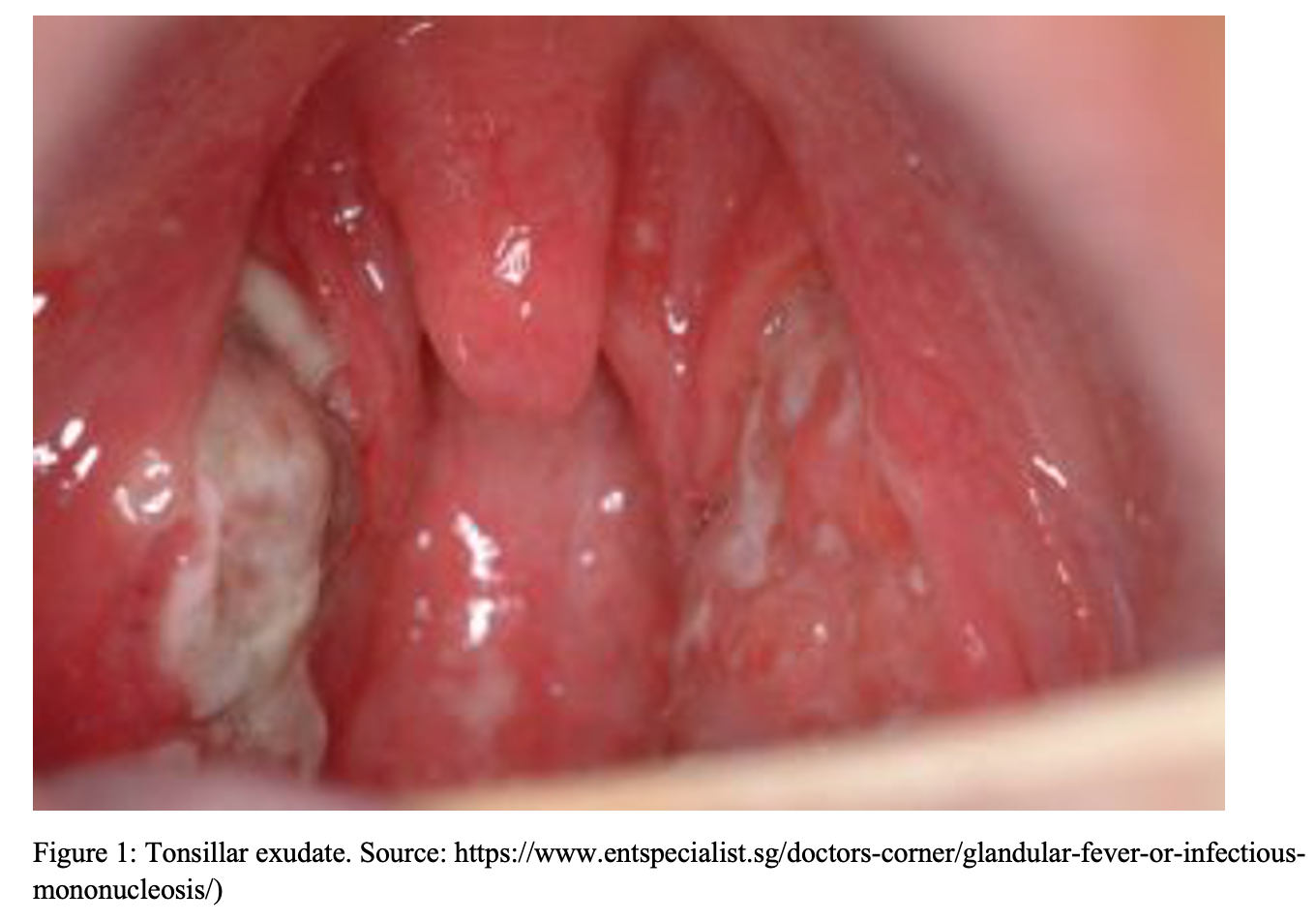
A 17-year-old male with history of asthma presents to the ED with a chief complaint of sore throat for the past 3 days, associated with decreased oral intake. He denies sick contacts. Vital signs include Temp 100.6F, BP 124/78, HR 92, RR 16, 96% on RA. On exam the patient has swollen, erythematous tonsils with grayish exudate, as well as enlarged, tender anterior and posterior cervical chain lymph nodes.
What is the most likely diagnosis?
Answer: Infectious mononucleosis [1-14]
Etiology
- Epstein-Barr Virus (EBV) is responsible for the vast majority (90%) of Infectious mononucleosis (IM) cases
- EBV spreads through contact, typically via salivary secretions
- Toy and utensil sharing in young children and kissing in adolescents are thought to be primary modes of transmission
- Oral shedding persists for a median duration of 6 months after illness onset [1]
- Infectious mononucleosis is a syndrome marked by pharyngitis, cervical lymphadenopathy, and fever. Not all people who contract EBV will have the full syndrome or be symptomatic.
- Roughly 10% of individuals with IM will not have EBV as the causative agent [2]
- Other viruses that may cause IM include CMV, HIV, Adenovirus, Hepatitis A, Toxoplasma, and Rubella
Epidemiology
- Estimated 90-95% of people worldwide become seropositive for EBV in their lifetime [3]
- Clinical prevalence of Infectious Mononucleosis (IM) peaks between 15 and 24 years of age [4]
- The disease does not display seasonality [5]
- A prospective study of primary EBV infection in college freshmen demonstrated 89% with symptomatic infection: 77% with full IM syndrome and 12% with atypical symptoms [1]
- While primary EBV infection is common in young children, it is most often clinically asymptomatic, with <10% developing symptoms
- Differences in children and young adults regarding symptomatic infection are not fully understood, the size of viral inoculum at time of infection is considered a possible factor
Pathophysiology
- Initial spread via salivary secretions
- Once the virus has infected B cells in lymphoid areas in the oropharyngeal epithelium, it disseminates to the bloodstream [7]
- EBV-infected B cells then disseminate infection throughout the lymphoreticular system, causing enlargement of lymphoid tissues
- Primary EBV infection of B lymphocytes induces an immunologic response in the form of circulating antibodies directed against the viral antigens and other unrelated antigens, including those found on sheep and horse red cells (heterophile antibodies) [8]
- This immunologic response is likely responsible for the clinical manifestations of IM
Clinical Features

- The most common signs and symptoms of IM include fever, pharyngitis, lymphadenopathy, and fatigue
- Physical exam may show pharyngeal inflammation, tonsillar exudates that are white, grey/green, or even necrotic appearing, and palatal petechiae
- Palatal petechiae has 95% specificity for IM in patients with sore throat [9]
- Splenomegaly seen in 7-53% of patients with IM [9]
- Splenic rupture is a rare but potentially fatal complication of IM
- Estimated prevalence of 1-2 ruptures per thousand cases of IM [10]
- Rupture most common in men who are <30 years old, with 84% occurring within 4 weeks of symptom onset[11]
- Systematic review demonstrated average time of rupture to be 14 days after symptom onset [11]
- Rupture is spontaneous in 50% of cases
- Clinical variants of IM exist, including a “typhoidal form” which involves fever and lymphadenopathy without pharyngitis
- These cases may be heterophile antibody negative
- Possible causes of heterophile antibody negative IM include CMV, acute HIV, and toxoplasmosis
- The most important viral infection to differentiate from IM is acute HIV infection, as earlier diagnosis of HIV can make a significant difference in the outcomes for patients
- abrupt onset of symptoms, including diarrhea or pharyngeal edema without associated tonsillar exudate or hypertrophy are features that help to distinguish acute HIV from EBV mononucleosis [12]
- mucocutaneous ulcerations also help distinguish acute HIV from EBV mononucleosis.
Evaluation
Physical Examination
- Begin with evaluation of airway, breathing and circulation
- Evaluate face and exposed skin for presence of morbilliform rash
- Examine for cervical, axillary, and inguinal lymphadenopathy
- Presence of posterior cervical, axillary, or inguinal adenopathy increase likelihood of IM diagnosis [9]
- Examine pharynx for tonsillar hypertrophy or exudates, inspect palate for presence of petechiae
- Examine abdomen for presence of splenomegaly or abdominal pain to palpation
Labs
- Labs that may assist with evaluation for IM include:
- Elevated WBC, which may reveal increased lymphocyte count or presence of atypical lymphocytes
- The presence of greater than 10% atypical lymphocytes significantly increases the likelihood of mononucleosis, especially when accompanied by lymphocytosis [9]
- A self-limited rise in LFTs is seen in the majority of patients with IM
- When coupled with pharyngitis abnormal LFTs strongly suggest IM
- Routine testing for LFT elevation is not indicated, and there is not a need to trend LFTs in immunocompetent patients with IM [13]
- Elevated WBC, which may reveal increased lymphocyte count or presence of atypical lymphocytes
DDX
- Cytomegalovirus infection, Toxoplasmosis, Strep pharyngitis, Neisseria gonorrhoeae, adenovirus infection, human immunodeficiency virus infection, acute leukemia [3]
Diagnosis
- Diagnosis can be suspected based on clinical exam findings listed above
- Heterophile antibody test or EBV specific antibody testing should be done to confirm diagnosis
- Heterophile antibody testing may be falsely negative early in clinical course, as high as 25% falsely negative during first week of symptoms
- Repeat testing or EBV-specific antibodies should be pursued when strong clinical suspicion exists
- Diagnostic testing especially important in pregnant patients as CMV, HIV, and toxoplasma can have significant adverse effects [3]
- Confirming the diagnosis of EBV IM is helpful in that patients can be informed of rare complications of IM such as splenic rupture and airway obstruction. It can also help explain a prolonged course of fatigue.
Treatment
- Supportive care is the mainstay of primary EBV Infectious mononucleosis
- NSAIDs (ibuprofen 400 mg PO q4-6h prn) or Acetaminophen (325mg PO q4-6h prn) for fever, pharyngitis, malaise
- Adequate hydration and nutrition
- Use of corticosteroids are not typically recommended in IM except for certain indications, such as impending airway closure, or other severe/rare conditions such as aplastic anemia or fulminant liver failure
- Antivirals such as acyclovir have not been shown to have clinical benefit and should not be routinely used [14]
- Given the risk of splenic rupture, all patients should be advised to avoid contact sports or other similar activities for 4 weeks after symptom onset
- Consider ultrasound of the spleen prior to return to play
Disposition
- Patients are typically safe for discharge home with outpatient follow up and return precautions
Summary and Pearls
- If a patient presents with mononucleosis-like syndrome but has abrupt onset of symptoms, diarrhea, or mucocutaneous lesions, consider HIV work up
- Axillary, inguinal, or posterior cervical adenopathy, palatal petechiae, and splenomegaly greatly increase likelihood of mononucleosis in the correct clinical setting
- Splenic rupture is a rare but potentially fatal effect of IM. The greatest risk is in males under the age of 30, within 4 weeks of symptom onset. Avoid contact sports for 4 weeks and give return precautions for any new-onset abdominal pain
- False-negative rate of heterophile antibody testing can be as high as 25% in first week of symptoms, consider repeat test or EBV specific antibody testing, especially for pregnant patients
Further Reading:
https://wikem.org/wiki/Infectious_mononucleosis


