Authors: Victoria Serven, MD (EM Resident Physician, Carolinas Medical Center, Charlotte, NC); Bryant Allen, MD (@bryantkallen, EM Attending Physician, Carolinas Medical Center, Charlotte, NC) //Reviewed by: Alex Sheng, MD, MHPE (@TheShenger); Alex Koyfman, MD (@EMHighAK); Brit Long, MD (@long_brit)
Case 1: A 23-year-old male presents with a single stab wound to the left groin. The blade was estimated to be 3-4 inches long. The wound is 2 cm wide with dark blood slowly oozing from the site. Vital signs as follows: BP 153/75, HR 92, RR 12, SpO2 98%. What is the next step in management? How would you evaluate for this patient’s injuries?
Case 2: A 27-year-old male with penetrating ballistic wound to the right groin. The right thigh is firm, dorsalis pedis pulse is absent on the right and 2+ on the left. Vital signs as follows: BP 140/80, HR 114, RR 16, SpO2 99%. What is your biggest concern given this patient’s physical exam findings? What is your next step in management?
Case 3: A 25-year-old male with penetrating ballistic wound to the right groin. The patient is agitated and uncooperative with exam. The abdomen is firm and rigid. Vitals signs as follows: BP 92/47, HR 146, RR 32, SpO2 96%. What is your next step in management?
Anatomy
The groin is the border to the box—the line between lower extremity and the trunk. The superior landmark of the groin follows the inguinal ligament between the anterior superior iliac spine (ASIS) and the pubic tubercle. The medial border is an artificial line which begins at the pubic tubercle separating the adductor muscles, which lie medial to the border, from the pectoralis and iliopsoas muscles. The lateral border runs distally from the ASIS to meet the medial order near to middle portion of the rectus femoris muscle thusly completing the triangular region we recognize as the groin (1).
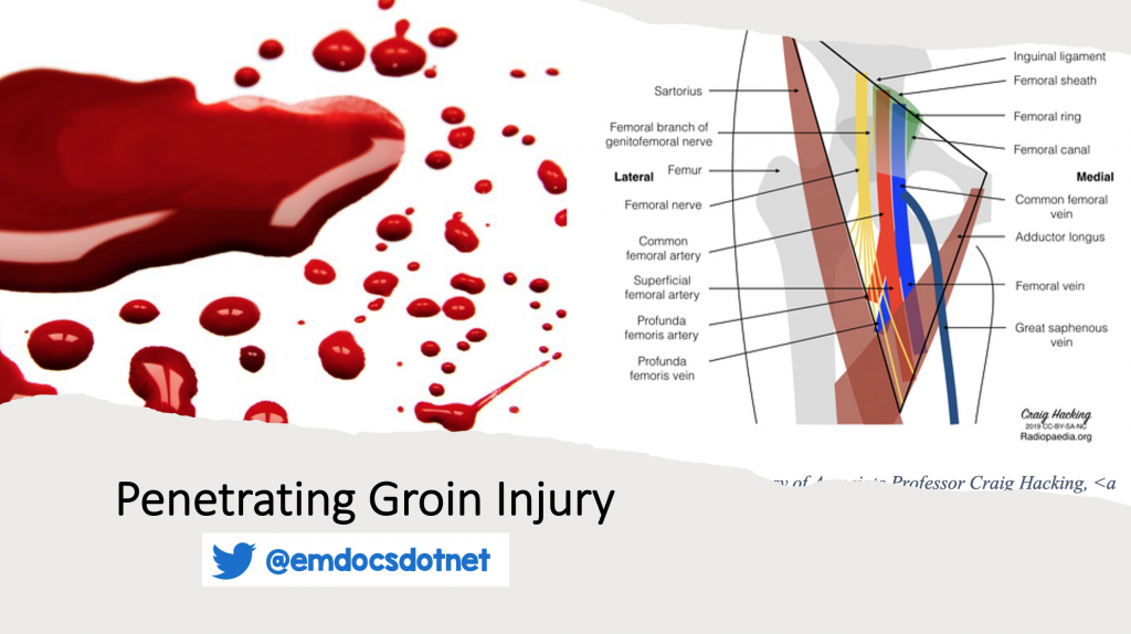
 While this area contains several large muscles important for the function of the lower extremity, in regard to the trauma patient, we are most concerned with neurovascular structures contained within the femoral sheath which lies within the boundaries of the groin. The femoral sheath is an extension of transverse facia anteriorly and iliac fascia posteriorly and therefore provides an area of communication between the abdominal cavity and the groin (2). Its location can be further delineated as located within the femoral triangle, which is marked by the inguinal ring superiorly, adductor longus muscle medially, and the sartorius muscle laterally. The contents of the femoral sheath from lateral to medial include the femoral nerve, femoral artery, and femoral vein. Just medial to the femoral vein you will find the proximal portion of the great saphenous vein which empties into the femoral vein (2). Medical students often learn the contents of the femoral sheath with the mnemonic NAVEL—again from lateral to medial the letters stand for Nerve, Artery, Vein, Empty (femoral canal), and Lymph node.
Approach to the Patient with Penetrating Groin Trauma
Penetrating trauma can be broken down to three basic mechanisms (3):
- Low velocity i.e. stab wounds
- Medium velocity projectile i.e. hand guns
- High velocity projectile i.e. explosive devices, rifles, and high-powered weapons such as those used by the military
One of the first steps in assessing severity of injury in patients with penetrating wounds to the groin will be to look for signs of vascular injury. Just as in other areas of the body, hard signs of vascular injury to the groin include (4):
- Active hemorrhage
- Expanding Hematoma
- Distal ischemia
- Bruit or thrill
- Pulse deficit
Patients with hard signs of vascular injury or those who are hemodynamically unstable will need to go to operating room (OR) for definitive management of their injuries. For these patients efforts in the trauma bay should be focused on hemorrhage control, resuscitation, and stabilization in a timely manner to facilitate expeditious transport to the OR. Achieving hemostasis through direct pressure may be difficult as injured vessels may be hidden deep under subcutaneous tissue or extend into the abdominal cavity (2). Furthermore, hemorrhage should be controlled at both the proximal and distal ends of the injured vessel. Techniques for controlling bleeding are discussed below.
Once hemorrhage is controlled, patients should undergo a thorough secondary evaluation to assess for additional injuries. An abdominal exam should be preformed to assess for peritonitis which may be a result of bowel or genitourinary (GU) system injury (5). Blood at the meatus may suggest urethral or bladder damage. A rectal exam should be performed to assess for bleeding that may result from bowel penetration. Ultrasound can be used to quickly assess for free fluid in the abdominal cavity as part of a focused assessment with sonography in trauma (FAST) exam. If there is any concern for pelvic or intra-abdominal injuries in the stable patient, computed tomography (CT) imaging should be performed. Imaging with rectal contrast can help identify rectal or colonic injuries (5). CT cystogram or urogram should be performed if GU injuries are suspected (6). Xray can be useful both in identifying bony injuries and the location of any retrained foreign bodies (7).
If the patient remains hemodynamically stable following the secondary survey, a more thorough exam should be performed on the lower extremity to assess for more subtle injuries. A complete sensory and motor exam will aid in identifying injuries to the femoral nerve. Signs of injury include (8):
- Weakness with hip flexion
- Weakness with knee extension
- Numbness over medial thigh
- Numbness over calf
- Reduced patellar reflex
The absence of hard signs of vascular injury described above does not rule out a more subtle injury such as formation of a pseudoaneurysm or arteriovenous fistula. Concerning soft signs for a more occult arterial injury include (9):
- Neurologic Deficit
- Palpable Pulse Deficit
- Nonexpanding hematoma
- Variation in doppler signal between extremities
- Abnormal Ankle-Brachial Indexes (ABIs) <0.9
If there is any concern for vascular injury, and the patient is stable, ABIs should be performed. Abnormal ABIs or presence of any of the other soft signs listed above requires further investigation with CT angiography of the lower extremity (gold standard) or duplex ultrasound (if CT is not available). In patients without hard or soft signs, normal ABIs, and/or normal imaging vascular injury can be safely ruled out and the patient discharged (9).
Hemorrhage Control
Trauma to the groin and axilla can be described as “junctional injuries” as they represent a transition area between the limb and the trunk. As described previously, the groin contains the proximal femoral vessels and injury to these vascular structures can lead to brisk exsanguination. While distal bleeding can be controlled with a tourniquet, control of the proximal portion is often difficult as it relies on tamponade of the truncal structures. In this section we will review several of the most popular techniques for gaining control of proximal vessel hemorrhages (10).
External Compression of Damaged Vessel
Traditional methods of management focus on occluding the vessel by deep wound packing followed by application of a pressure dressing plus or minus external compression of the abdominal or pelvic vessels (11).
There are now several types of gauze on the market that are interlaced with chemical hemostatic agents to aid in coagulation. Such agents include QuikClot, Combat Gauze, HemCon, among others. Although these agents have been shown to reduce hemorrhage when compared to traditional gauze; they often fail to provide significant source control in the setting of severe bleeds including deep penetrating wounds or traumatic amputations (12). Thus, it is important to have other tools and techniques in mind when attempting to address these injuries.
Injectable Hemostatic Sponges include such commercial devices as the XSTAT and are essentially large syringes filled with small sponges made of cellulose which expand when they come in contact with liquids. These small sponges can be injected into wounds where they would ideally expand and provide enough compression to restrict or stop blood flow from the damaged vessel. These devices have been used by the military for several years and are beginning to gain popularity in civilian medicine (12).
Similarly, the brand Celox offers a device to inject hemostatic granules into a wound. Floseal is acts along the same principles to inject a gelatin matrix crosslinked by glutaraldehyde and a thrombin solution (1000 U/ml). These agents may aid in clot formation directly at the sight of application which is independent of the patient’s intrinsic clotting cascade (12).
Wound Clamps, such as the iTClamp, are portable direct mechanical pressure devices which are deployed at the sight of injury to provide wound closure which ideally will stop exsanguination by allowing a hematoma to form which will in time provide enough pressure to tamponade the vessel. These devices were specifically designs to address junctional bleeding. Though only a limited number of studies have been performed using this device, it has shown increased rates of hemostasis when combined with wound packing either with gauze or hemostatic sponges (11).
In addition to local control of hemorrhage, efforts should be made to occlude bleeding from a more proximal source. In the instance off penetrating trauma to the groin this mean occlusion of the iliac artery or abdominal aorta.
If there is a spare provider assisting in the resuscitation or responding to the initial scene it may be worth your efforts to instruct them to use their body weight to compress the intraabdominal structures. Several studies have been performed to identify the most effective location, technique, and amount of weight needed to achieve hemostasis with external compression of these vessels. Blaivas et al used ultrasound to determine how much weight would be required to occlude flow to the common femoral artery (13). They found that velocity of flow began to decrease with as little at 20 pounds of pressure applied to the abdominal aorta but to completely occlude flow in most individuals required ~105 pounds (range of 80 – 140 pounds). Absence of flow was achieved with only 82 pounds (range of ~60 – 100 pounds) with application of pressure to the proximal iliac artery. Volunteers were unable to tolerate enough pressure to achieve occlusion with application to the distal iliac artery, interestingly initial application of pressure over this area actually increased the velocity of flow through the vessel. Possible techniques for compression include placing a knee over the vessel or two locked arms over a fist (14). Other studies have found that the amount of pressure a rescuer could provide was directly proportional to their body weight and that the knee method allowed for greater pressure than the fist method (15).
There are external compression devices designed to produce similar outcomes as direct compression by an individual. An example of one such device is the Abdominal Aortic and Junctional Tourniquet. Advantages of this device include reduction in personnel required to be transported with the patient, allows for greater pressure (up to 300 mmHg) possibly more reliable continuous application of pressure, allows more space at the groin for additional treatments or procedures (16). One study using swine models found similar rates of hemostasis when compared to REBOA (17). This device is light weight and can be deployed in approximately 1 minute. Just as with an extremity tourniquet this device can cause ischemia and reperfusion injury to distal tissues and it should be left in place for as short of a time as possible (18).
Internal Compression of Proximal Vessel
If you can identify the bleeding vessel a simple but effective option may be to inflate a Foley catheter within the offending structure. This has been shown in several case studies to be an effective method to obtain hemostasis in both groin and neck penetrating trauma (19, 20).
Resuscitative Endovascular Balloon Occlusion of the Aorta (REBOA) is another option for obtaining temporary internal hemostasis. For the purposes of REBOA catheter placement, the aorta is divided into 3 zones: Zone 1 extends from the left subclavian artery takeoff down to the level of the celiac artery; Zone 2 extends from the celiac artery to the renal arteries; and Zone 3 extends from the renal arteries caudally to the aortic bifurcation. Access should be obtained by an experienced provider via the uninjured femoral artery and after insertion of the catheter, the balloon can be deployed in Zone 3 or the distal abdominal aorta which will impede blood flow to the iliac vessels (21). Use of REBOA remains controversial as there is no evidence showing that it improves mortality and may result in serious complications such as ischemia to distal structures including the spinal cord or direct vessel injury. For now, it is recommended that only centers with expedient access to a means of definitive surgical management utilize these techniques. The target time to device removal should be within 30 minutes with a maximal time of 60 minutes (21).
Case Conclusion
Case 1: The patient is stable so a complete secondary survey should be performed looking for hard and soft signs of vascular injury. If any hard signs are present, the patient will need to go to the OR. If there are soft signs or an ABI <0.9, the patient will need to undergo advanced imaging in the form of a CTA. If there are not hard or soft signs and the patient has an ABI >0.9 the likelihood of a vascular injury is very low.
Case 2: The patient has hard signs of vascular injury and needs to go to the OR. His vital signs are stable for now. The focus should be on hemostasis and expeditious transfer to the operating theater.
Case 3: The patient is unstable and showing signs of an intraabdominal injury which may include trauma to the blood vessels, bowel, or bladder. The focus should be on stabilizing the patient as quickly as possible through external or internal occlusion of the damaged artery as this patient is at risk for exsanguinating before reaching definitive management in the OR.
Key Points
- Hard signs of vascular injury are an indication for immediate operative
- If there is any concern for vascular injury, preform ABIs followed by CT angiogram of the lower extremities.
- In times of major hemorrhage when the OR is not immediately available, efforts should be focused on compressing the injured vessel to obtain hemostasis.
- Several commercial devices are available to help aid in providing direct pressure over the injury vessel.
- If the injured vessel cannot be reached, applying pressure to the abdominal aorta can slow the rate of blood loss. Most adults will require around 100 pounds of pressure to occlude flow.
- A REBOA device deployed in Zone 3 may be used to attempt internal occlusion of a proximal vessel.
References
- Falvey EC, Franklyn-Miller A, McCrory PR. The groin triangle: a patho-anatomical approach to the diagnosis of chronic groin pain in athletes. British Journal of Sports Medicine 2009;43:213-220.
- Chaurasia, BD, Garg, K, Mittal PS, Chandrupatla M. BD Chaurasia’s Human Anatomy: Regional and applied, dissection and clinical, 7th New Delhi: CBS Publishing; 2017.
- Kuhajda I, Zarogoulidis K, Kougioumtzi I, et al. Penetrating trauma. Journal of Thoracic Disease 2014; 6(Suppl 4):S461–S465. https://doi.org/10.3978/j.issn.2072-1439.2014.08.51
- Dennis JW, Frykberg ER, Crump JM, et al. New perspectives on the management of penetrating trauma in proximity to major limb arteries. Journal of Vascular Surgery 1990;11(1):84-93. doi:10.1067/mva.1990.16942
- Dreizin D, Boscak AR, Anstadt MJ, et al. Penetrating Colorectal Injuries: Diagnostic Performance of Multidetector CT with Trajectography. Radiology 2016;281(3):749-762. doi:10.1148/radiol.2015152335
- Haroon SA, Rahimi H, Merritt A, et al. Computed tomography (CT) in the evaluation of bladder and ureteral trauma: indications, technique, and diagnosis. Abdom Radiol 2019;44(12):3962-3977.
- Frykberg ER, Dennis JW, Bishop K, et al. The reliability of physical examination in the evaluation of penetrating extremity trauma for vascular injury: results at one year. J Trauma 1991;31(4):502-11. doi: 10.1097/00005373-199104000-00009. PMID: 2020036.
- Moore AE, Stringer MD. Iatrogenic femoral nerve injury: a systematic review. Surg Radiol Anat. 2011 Oct;33(8):649-58. doi: 10.1007/s00276-011-0791-0. Epub 2011 Feb 17. PMID: 21328076.
- deSouza IS, Benabbas R, McKee S, et al. Accuracy of Physical Examination, Ankle-Brachial Index, and Ultrasonography in the Diagnosis of Arterial Injury in Patients With Penetrating Extremity Trauma: A Systematic Review and Meta-analysis. Acad Emerg Med 2017;24(8):994-1017. doi: 10.1111/acem.13227. PMID: 28493614.
- van Oostendorp SE, Tan EC, Geeraedts LM Jr. Prehospital control of life-threatening truncal and junctional haemorrhage is the ultimate challenge in optimizing trauma care; a review of treatment options and their applicability in the civilian trauma setting. Scand J Trauma Resusc Emerg Med 2016;24(1):110. doi: 10.1186/s13049-016-0301-9. PMID: 27623805; PMCID: PMC5022193.
- Stuart S, Zarow G, Walchak A, et al. Pilot Study of a Novel Swine Model for Controlling Junctional Hemorrhage Using the iTClamp in Conjunction With Hemostatic Agents. Military Medicine 2019;184:367-373. doi: 10.1093/milmed/usy337.
- Peng HT. Hemostatic agents for prehospital hemorrhage control: a narrative review. Military Medical Research 2020;7(1):13. https://doi.org/10.1186/s40779-020-00241-z
- Blaivas M, Shiver S, Lyon M, Adhikari S. Control of hemorrhage in critical femoral or inguinal penetrating wounds–an ultrasound evaluation. Prehosp Disaster Med 2006; 21(6):379-82. doi: 10.1017/s1049023x00004076. PMID: 17334183.
- O’Dochartaigh D, Picard CT, Brindley PG, Douma MJ. Temporizing Life-Threatening Abdominal-Pelvic Hemorrhage Using Proprietary Devices, Manual Pressure, or a Single Knee: An Integrative Review of Proximal External Aortic Compression and Even “Knee BOA”. J Spec Oper Med 2020;20(2):110-114. PMID: 32573746.
- Douma M, Brindley PG. Abdominal aortic and iliac artery compression following penetrating trauma: a study of feasibility. Prehosp Disaster Med 2014;29(3):299-302. doi: 10.1017/S1049023X1400051X.
- Croushorn J. Abdominal aortic and junctional tourniquet controls hemorrhage from a gunshot wound of the left groin. J Spec Oper Med 2014;14(2):6-8.
- Schechtman DW, Kauvar DS, De Guzman R, et al. Abdominal aortic and junctional tourniquet versus zone III resuscitative endovascular balloon occlusion of the aorta in a swine junctional hemorrhage model. J Trauma Acute Care Surg 2020;88(2):292-297. doi: 10.1097/TA.0000000000002553.
- Kheirabadi BS, Terrazas IB, Miranda N, et al. Long-term consequences of abdominal aortic and junctional tourniquet for hemorrhage control. J Surg Res 2018; 231:99-108. doi: 10.1016/j.jss.2018.05.017.
- Jose A, Arya S, Nagori SA, Thukral H. Management of Life-Threatening Hemorrhage from Maxillofacial Firearm Injuries Using Foley Catheter Balloon Tamponade. Craniomaxillofacial Trauma & Reconstruction2019;12(4):301-304. doi:1055/s-0039-1685461
- Singh R, Trickett R. Balloon tamponade with a Foley catheter for extremity stab injury. Injury Extra 2010;41(12), 202. doi:10.1016/j.injury.2010.07.184
- Bulger EM, Perina DG, Qasim Z, et al. Clinical use of resuscitative endovascular balloon occlusion of the aorta (REBOA) in civilian trauma systems in the USA, 2019: a joint statement from the American College of Surgeons Committee on Trauma, the American College of Emergency Physicians, the National Association of Emergency Medical Services Physicians and the National Association of Emergency Medical Technicians. JTrauma Surg Acute Care Open 2019;4(1):e000376. https://doi.org/10.1136/tsaco-2019-000376


