Today on the emDOCs cast with Brit Long (@long_brit), we cover orbital cellulitis and how to differentiate it from periorbital cellulitis.
Episode 151: Orbital Cellulitis
Background:
- Orbital cellulitis:
- Inflammation of the globe contents and surrounding soft tissues behind the orbital septum.
- Usually due to an infection.
- Can be vision-threatening.
- Preseptal cellulitis (a.k.a. periorbital cellulitis):
- Preseptal cellulitis is a different disease than orbital cellulitis.
- It is in a different anatomic location and does not carry the same morbidity and mortality as orbital cellulitis.
- Before antibiotics vision loss was common, and mortality was 20-40%.
- Current mortality is ~1% with timely diagnosis and treatment.
Epidemiology:
- Orbital cellulitis is 10 times more common in kids than adults.
- Orbital cellulitis occurs more often in the winter and spring because URIs are more common at that time of year.
- Sinusitis is the most common cause of orbital cellulitis.
- It most commonly involves the ethmoid sinus in kids <7 years old (as that is the only sinus developed at that age) and the frontal or multiple sinuses in older children and adults.
- Other causes include dacryocystitis, dental infections, and trauma.
- Haemophilus influenzae type B was previously the most common causative organism. Staphylococcus aureus and Streptococcus species are more common now.
- MRSA accounts for ~5% of cases.
- Gram-negative microbes and anaerobes are also possible, most commonly caused by dental infections.
- Fungal infections like mucormycosis and Aspergillosis are possible in immunocompromised patients such as patients with HIV or neutropenia.
Anatomy:
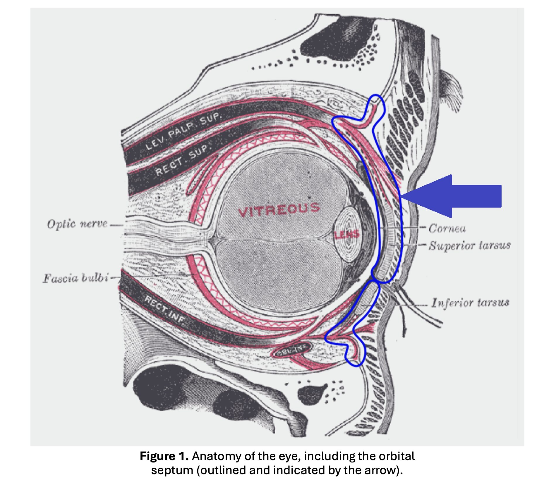
- The orbital septum is a fibrous membrane that acts as the boundary between the anterior orbit and the posterior orbit.
- In front of the septum (“preseptal”) are the eyelids.
- Posterior to the septum (“postseptal”) are the orbital contents, the retina, the optic nerve, and the vasculature that drains the orbits. This is what is affected by orbital cellulitis.
- Some of the veins in this area do not have valves, allowing an infection to track from the orbits to the cavernous sinus or the brain, resulting in meningitis or intracranial abscess.
Presentation:
- 80% of orbital cellulitis cases can be accurately diagnosed clinically.
- A painful, red eye is the most common presentation.
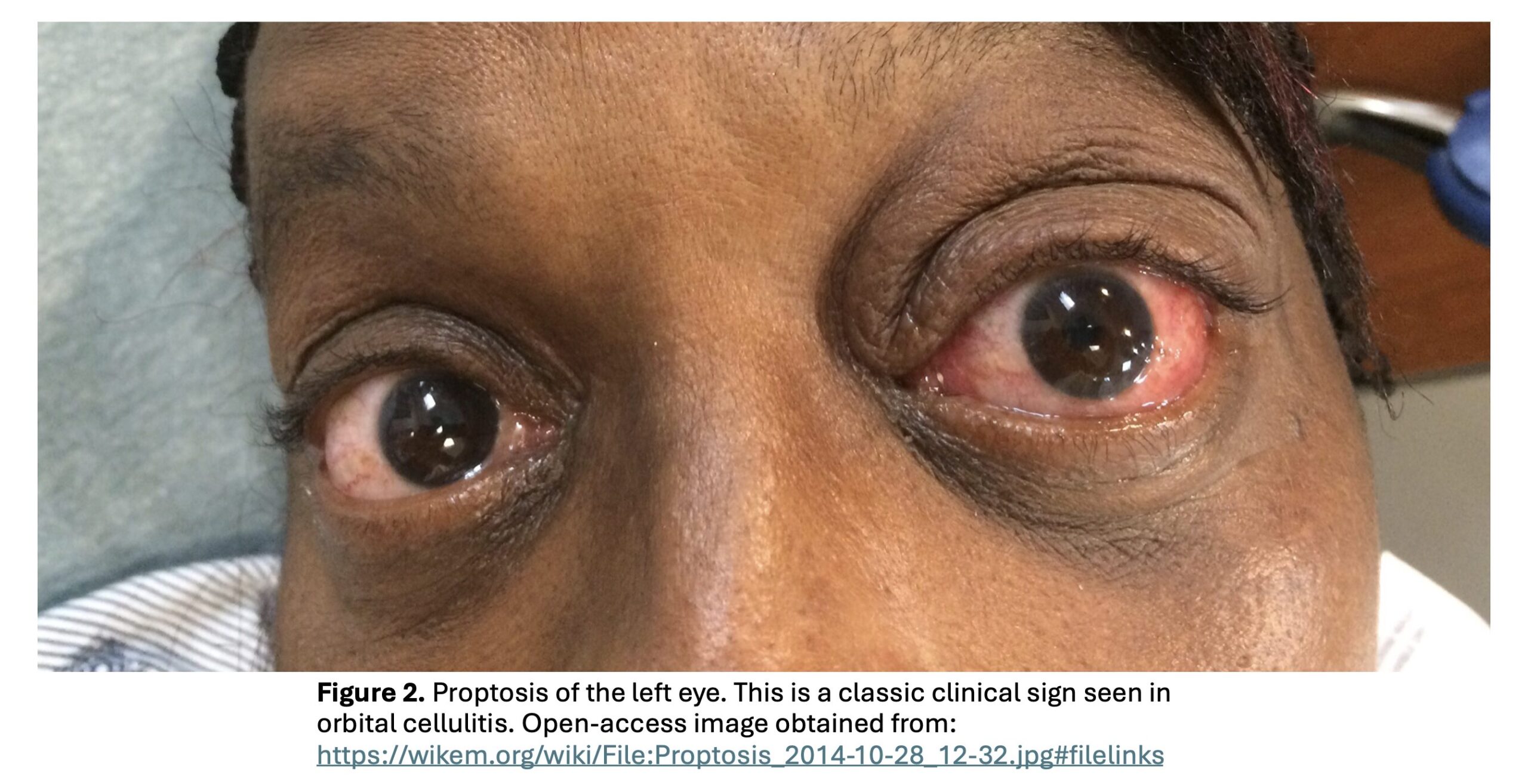
- Over 90% of patients will have eyelid swelling. 80% have periorbital/eyelid erythema.
- Restricted eye movements present in 65-100% of cases, pain with eye movement 33-55%, proptosis 8-45%, photophobia 20%, diplopia 15%, change in vision up to 20%.
- No clinical finding is specific.
- Red Flag findings for orbital cellulitis:
- Pain with eye movement
- Restricted eye movement
- Vision changes or changes in color vision
- Diplopia that improves with closing the affected eye
- Relative afferent pupillary defect (RAPD)
- Proptosis
- Try standing over the patient and looking down over the forehead and eyes to pick up more subtle cases of proptosis.
- Consider in any patient with URI symptoms AND eye symptoms or dental symptoms AND eye symptoms.
- Ocular compartment syndrome (OCS) can also occur due to inflammation/infection, so be sure to check intraocular pressure if you have concerns for orbital cellulitis.
- OCS is much less common in orbital cellulitis than in trauma with retrobulbar hemorrhage.
Diagnosis:
- Labs
- Labs cannot make or rule out the diagnosis. They also cannot differentiate between orbital cellulitis and preseptal cellulitis.
- Leukocytosis and elevations in CRP and ESR are common but should not be used to rule out the disease if normal.
- Blood cultures positive in 3% of patients.
- Consultants may still ask for the labs, however, so consider getting them as a part of the workup.
- Culture of an intraorbital abscess is the most helpful lab, but this is not something that will be obtained in the ED.
- Imaging
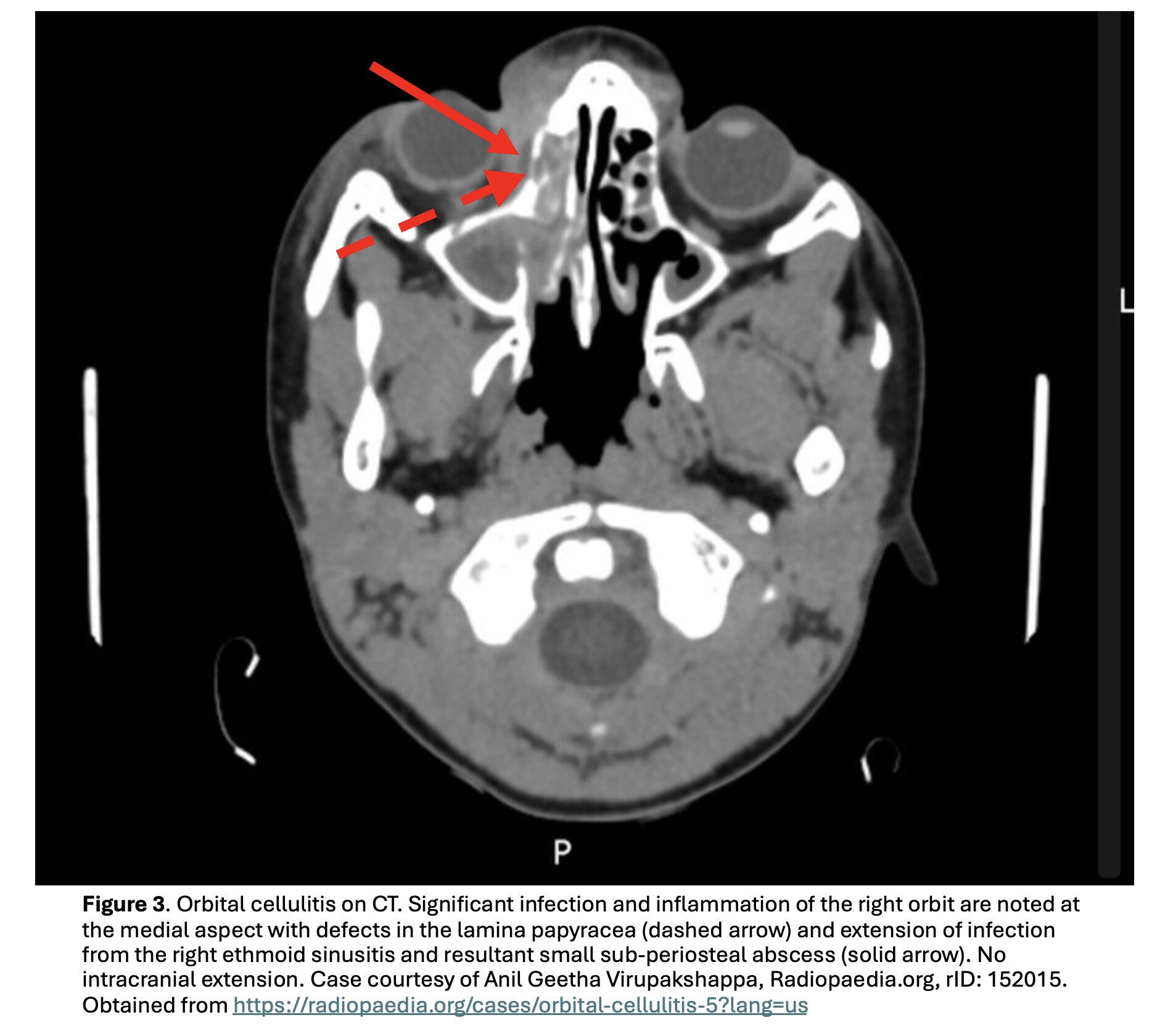
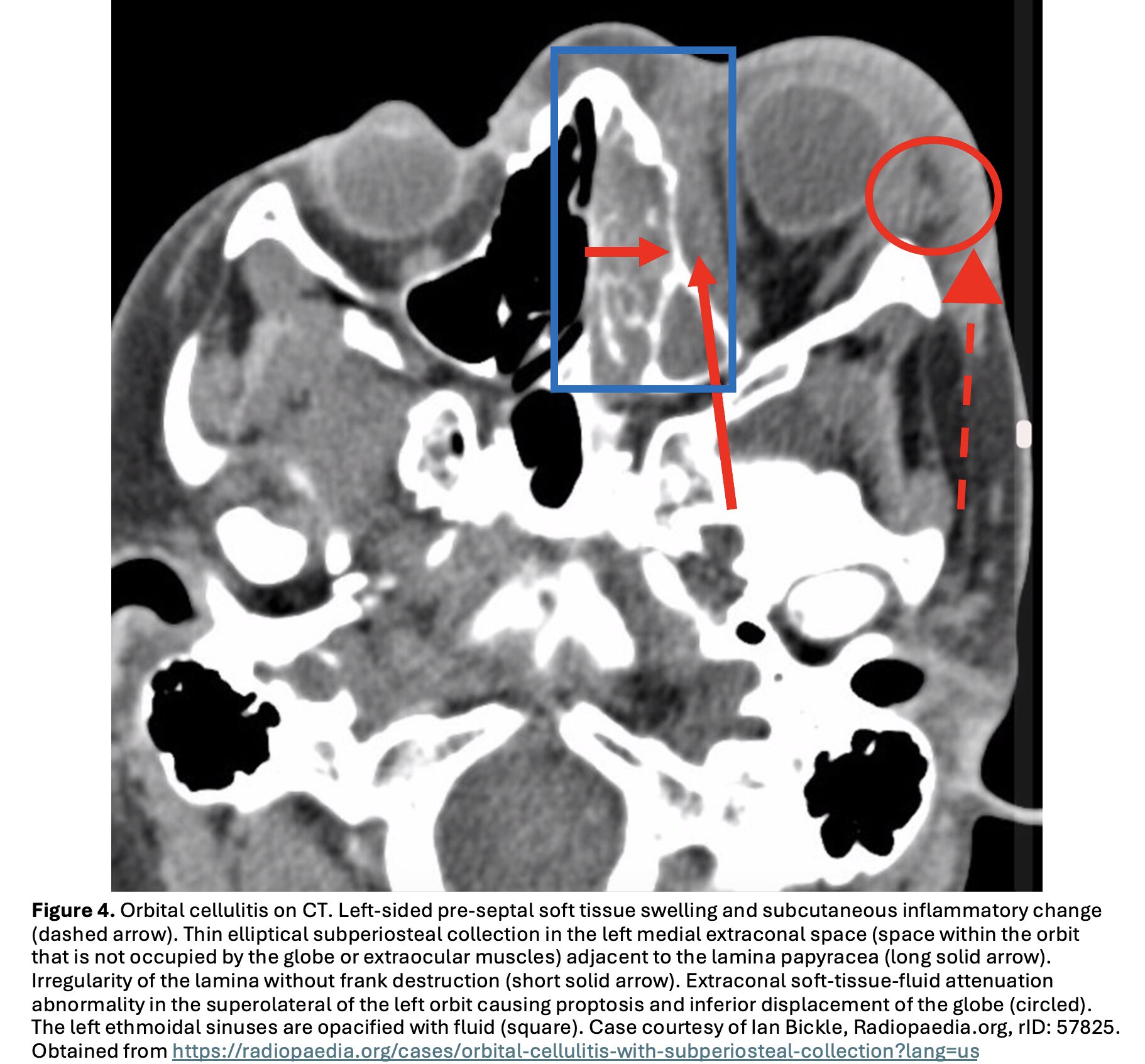
- CT of the brain and orbits with/without contrast is the go-to imaging modality.
- If there are any red flags, strongly consider obtaining CT.
- MRI may be better at finding soft tissue abnormalities.
- Consider MRI of the brain and orbits with/without contrast if CT is not diagnostic but you still have concerns.
- In pediatric patients, MRI can potentially be the first line, but this will depend on MRI availability.
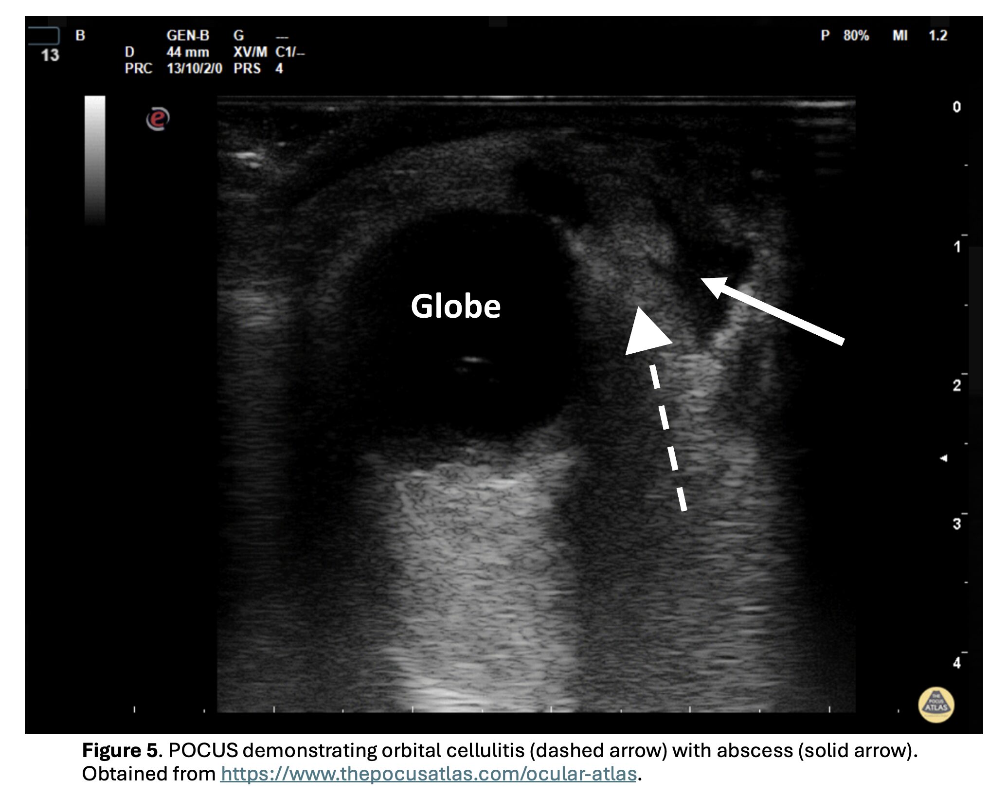
- Ultrasound has shown promise, but there is no definitive literature yet.
- Look for orbital edema, echogenic intraorbital fatty tissue, or an abscess.
- CT of the brain and orbits with/without contrast is the go-to imaging modality.
Treatment:
- Consult ophthalmology – patients may require surgical intervention.
- If there is intracranial involvement, neurosurgery consult is necessary.
- The faster these patients get to the specialist, the better.
- Antibiotics
- Cover staphylococcus, streptococcus, and anaerobes
- Good choices are vancomycin plus one of the following:
- Piperacillin-tazobactam
- Ampicillin-sulbactam
- Third generation cephalosporin
- Fluoroquinolone if penicillin allergic
- Steroids
- A 2021 Cochrane review found that steroids do NOT improve preservation of visual acuity, pain level on day 3, or duration of IV antibiotics.
- If the patient has increased intraocular pressure and concerned about OCS or they have severe pain or vision changes, they need an emergent lateral canthotomy and cantholysis – ideally within 2 hours. Vertical lid split is another option.
Pearls:
- Sinusitis is the most common cause of orbital cellulitis, but it should be considered in any patient with URI symptoms AND eye symptoms or dental symptoms AND eye symptoms.
- CT brain and orbits with/without contrast is the test of choice. Labs will not make or rule out the diagnosis.
- Red Flags include pain with eye movement, restricted eye movement, vision changes, diplopia that improves when the affected eye is closed, relative afferent pupillary defect (RAPD), or proptosis.
- Be sure to get an intraocular pressure measurement when red flags are present to evaluate for ocular compartment syndrome (OCS).
- Treatment includes antibiotics and early specialty consultation.
References:
- Tsirouki T, Dastiridou AI, Ibánez Flores N, et al. Orbital cellulitis. Surv Ophthalmol. 2018;63(4):534-553. PMID: 29248536
- Williams KJ, Allen RC. Paediatric orbital and periorbital infections. Curr Opin Ophthalmol. 2019;30(5):349-355.PMID: 31261188
- Gordon AA, Phelps PO. Management of preseptal and orbital cellulitis for the primary care physician. Dis Mon. 2020;66(10):101044. PMID: 32622679
- Fanella S, Singer A, Embree J. Presentation and management of pediatric orbital cellulitis. Can J Infect Dis Med Microbiol. 2011;22(3):97-100. PMID: 22942886
- Kornelsen E, Mahant S, Parkin P, et al. Corticosteroids for periorbital and orbital cellulitis. Cochrane Database Syst Rev. 2021;4(4):CD013535. Published 2021 Apr 28. PMID: 33908631
- McCallum E, Keren S, Lapira M, Norris JH. Orbital Compartment Syndrome: An Update With Review Of The Literature. Clin Ophthalmol. 2019;13:2189-2194. Published 2019 Nov 7. PMID: 31806931
- Ebright JR, Pace MT, Niazi AF. Septic thrombosis of the cavernous sinuses. Arch Intern Med. 2001;161(22):2671-2676. PMID: 11732931
- Pelletier J, Koyfman A, Long B. High risk and low prevalence diseases: Orbital cellulitis. Am J Emerg Med. 2023 Jun;68:1-9. PMID: 36893591