
Author: Lloyd Tannenbaum, MD (EM Attending Physician, APD, Geisinger Wyoming Valley, PA) // Reviewer: Brit Long, MD (@long_brit)
Welcome back to ECG Pointers, a series designed to make you more confident in your ECG interpretations. This week, we feature a post from Dr. Tannenbaum’s ECG Teaching Cases, a free ECG resource. Please check it out. Without further ado, let’s look at some ECGs!
Continued from last week (click here for last’s weeks case)
“Hello! Med Evac command? This is the hospital requesting a stat transfer for an unstable STEMI. 68-year-old male, inferior MI, briefly arrested but ROSC after 2 shocks. We have him on 2 different pressors and have given lytics as our cath lab is down. How soon can you get him out of here? I have an accepting team ready to do PCI about a 30-minute flight from here.”
“Doc, we are still mid transport to PICU. We cannot support your request at this time but will activate emergency procedures to try and get another helicopter team spun up as soon as possible. Best of luck to you and the team.”
“Understood, transfer center, get me EMS command please, we need to figure out how to get this patient out of here…” you say as you suddenly get cut off by Shannon.
“Doc, we need you back in the resus bay, the ectopy is increasing! I think he’s about to code again!” you hear her Shannon call out: “ATTENTION ATTENTION: PHARMACY TO THE RESUS BAY STAT! PHARMACY TO THE RESUS BAY STAT!” The hospital voice calls out.
“Stay on the phone with the transfer center and find someone, anyone with even a pickup truck that will transport this patient, please” you say to the clerk as you throw down the phone and run off with Shannon
“What’s going on in here?” you ask as you walk back into the resus bay.
“Ectopy is increasing, it looks like he’s going into a slow ventricular tachycardia. I stat-paged pharmacy to the bay for some anti-arrhythmics and …” the chief resident begins
“Wait. Slow Vtach? What?”
“Yeah!” says the chief resident. “Here’s his ECG, it looks like VT but the rate is less than 100, so a little weird. I just figured with his MI, he’s probably in VT and needs some anti-arrhythmics.”
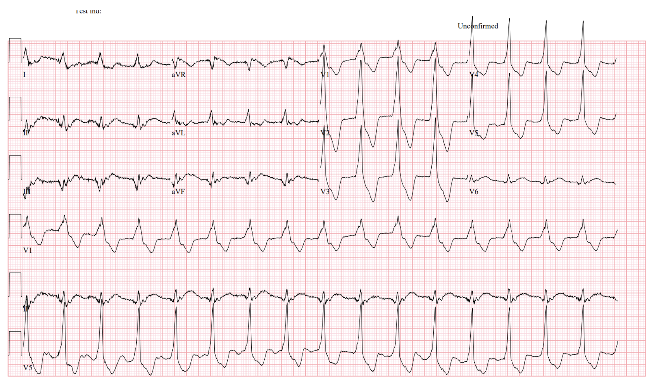
Rate: 95 beats per minute
Rhythm: Not sinus
Axis: Normal
Intervals: Wide complex QRS, no PR interval (no P waves) QTC looks mostly ok
Morphology: It sure looks like it could be VTach but the rate is too slow? Maybe?
Final read: Wide complex, regular rhythm with no P waves
“Hey guys, I came as quick as I could, what’s going on?” Scott the pharmacist asks.
“The STEMI-Lytics patient decided to go into VT. I’m going to need some amiodarone as soon as possible.”
The chief resident says. “Also, Shannon, can we get him hooked up to the defib pads too please?”
You, Shannon, and Scott look at each other and smile.
“WHAT?” the chief asks. “Why am I the only one concerned about VTach right now?”
“Well…” Scott begins.
“Come on, let me take this one. I’m the oldest in the room” you say.
“You’re not. But I’m going to let that go,” Shannon says. “You know I worked in the cardiac care unit for 12 years before I moved to the ER, right? I love it when new residents ran into this rhythm for the first time and lost their damn mind when they saw it, but it’s ok. I’ll let you do the big reveal.”
“Ok, Ok,” says Scott. “I have a bunch of medications to approve anyway. The floor is all yours.”
“You guys are so young,” you start, “you’ve always had a cath lab standing by, ready to take your patient away as soon as you call. But that wasn’t always the case. For a long time, lytics used to be the mainstay of treatment for patients with a STEMI. And with lytics, came reperfusion. Occasionally, you’d catch a slow, self-limited arrhythmia that looks like ventricular tachycardia, but it was too slow. This is AIVR, also known as Accelerated Idioventricular Rhythm or the reperfusion rhythm. In fact, take a look at this ECG and then I’ll tell you a story. This is the first time I saw AIVR, when I was an intern:”
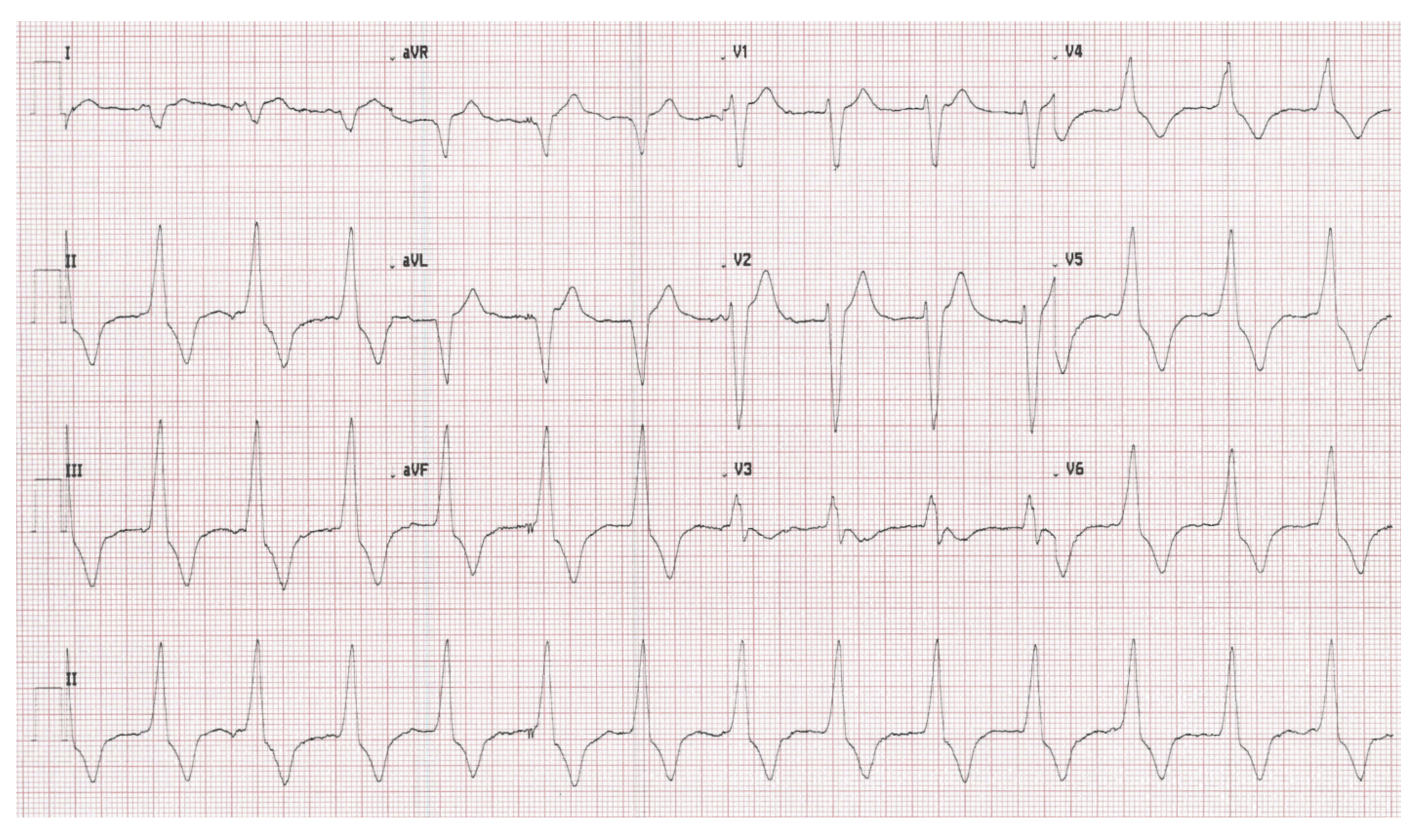
Rate: 80 beats per minute
Rhythm: Not sinus, no P waves
Axis: Right
Intervals: Wide QRS, no PR interval, QTc looks reasonable
Morphology: It looks a little bit like a left bundle at times, but not at others
Final read: This is another example of AIVR
“Back when some of us trained, our only option was to push lytics for an MI. And then, we’d see a weird, wide complex rhythm with no P waves that would occasionally have fusion or capture beats. We’d be worried about ventricular tachycardia, but the rate was usually under 100, making VTach unlikely.
The first time you’d see it, you’d have a small panic attack and reach for some kind of anti-arrhythmic, like amiodarone, but someone, nursing, pharmacy, or your attending, would stop you and tell you to just watch the tele for a bit and it’ll go away. The arrhythmia is often short lived and disappears as fast as it appeared. If you tried to convince the attending to give an anti-arrhythmic for this rhythm, they’d shoot you down and tell you it’s a benign rhythm that is a marker of reperfusion. In fact, treating this rhythm with anti-arrhythmics can cause precipitous drops in blood pressure and hemodynamic collapse! Usually, the best bet with AIVR is to leave the patient alone and the rhythm will resolve on its own.”
“So… We just do… nothing?” The chief asks.
“Correct,” says Shannon. “Unless you want this guy to code again?”
“No. Nope. No Thank you.” the intern stammers out. “I think we’ll go with it’s a benign rhythm and I’m going to walk away” he says as goes to check on his patients.
“I’m a little sad that you go to the do big reveal,” Scott tells you.
“Well. Being old has to come with some perks,” you reply. “Not just bottle of ibuprofen everywhere for when I sleep wrong…”
“Ok Grampa… Let’s get you back to work before you get cranky…” Scott says, rolling his eyes as you two head back to the main ER.
**This article does an excellent job of walking you through the history of AIVR and gives several additional examples of AIVR with capture and fusion beats. It’s definitely worth your time to take a look.**
Case Wrap Up
Just as walk back into the main ER, you get flagged down by your clerk.
“Doc! I have good news,” she yells to you. “The third Med Evac team is spun up and ready. They’ll be here in about 12 minutes to take this patient to a working cath lab. Send the nurse over to call report, we gotta get this guy out of here!”
Recap
1) AIVR is a transient rhythm often seen after given lytics in the setting of an MI.
2) This arrhythmia is often benign and does not require treatment. In fact, treating this rhythm with anti-arrhythmics can cause hemodynamic collapse!
3) The hallmark of AIVR is a rhythm that looks like VTach (wide complex, no P waves) but is too slow. Rates are usually between 60-100 beats per minute. Rates over 110 suggest Vtach. Rates under 50 are more consistent with a ventricular escape rhythm.