Introduction
- Incidence 2.5-3 per 100,000
- Peak incidence in 5th decade of life
- Significant cause of strokes in young/middle aged patients (5-22% of strokes in this age group)
Etiology/Predisposing Factors
- Spontaneous, minor trauma (e.g. sneezing, chiropractor manipulation, nose blowing, yoga), connective tissue disorders (e.g. Marfan’s, Ehlers-Danlos syndrome), genetic
- Underlying arteriopathy likely exists in patients with spontaneous dissection or due to minor trauma
- Major trauma: Elevated risk of carotid dissection with blunt or penetrating injuries involving the head, face, neck or thorax (and particularly in patients with skull base fractures, facial fractures, or TBI)
Pathophysiology
- Intimal tear or direct bleeding in arterial wall -> intramural hematoma -> stenosis of arterial lumen or aneurysm formation -> formation of associated thrombi which may embolize distally to cause ischemia
- If intracranial extension, may lead to subarachnoid hemorrhage
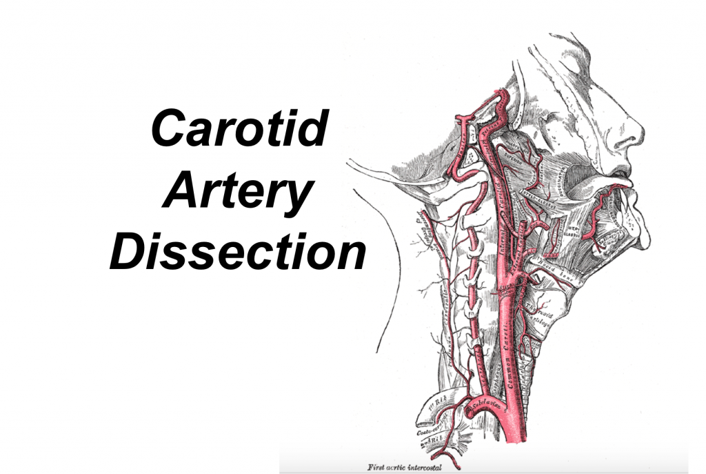
- Most commonly occurs in ICA, 2cm cephalad from bifurcation of common carotid to skull base
Signs and Symptoms
- Pain: Unilateral headache, neck pain, facial pain
- Partial horner’s syndrome (ptosis and miosis without anhidrosis); sympathetic fibers associated with facial sweating are associated closely with external carotid artery and NOT the ICA
- Cranial nerve palsies (CN XII most common), abnormal taste, pulsatile tinnitus
- Ischemia: TIA/stroke with associated anterior circulation deficits (hemiparesis, hemisensory loss, aphasia, neglect, amaurosis fugax)
- Patient with severe head/face/neck trauma that develops neurologic deficits
Diagnosis
- First line: CTA neck or MR angiography depending on local availability/practice pattern
- CTA generally used in patients with significant trauma
- Doppler ultrasound: Highly operator dependent, relatively poor sensitivity if low grade stenosis. Should NOT be used as first line test
- Angiography is traditional gold standard for diagnosis, but invasive and associated with complications
- Only if initial screening study is negative but high suspicion for carotid dissection
Management
- Spontaneous carotid dissection with evidence of acute ischemic stroke
- Recent meta-analysis shows similar safety in giving thrombolysis in dissection vs non-dissection related strokes (retrospective data, no RCTs)
- Thrombolysis CONTRAINDICATED if dissection is intracranial (risk of ICH) or involves the aorta (risk of aortic rupture)
- Antiplatelet vs anticoagulation
- Controversial; no published RCTs comparing either strategy (ongoing CADISS trial)
- Currently, no evidence of superiority in antiplatelet vs anticoagulation strategy. 2011 AHA/ASA guidelines state relative efficacy of either strategy is unknown
- Anticoagulation generally preferred if thrombus present in arterial lumen or severe stenosis
- Antiplatelet preferred if contraindication to anticoagulation, NIHSS >15, large infarct, intracranial extension of dissection
- No clear criteria for endovascular therapy (stent)
- Considered when new ischemic symptoms develop despite being on antiplatelet or anticoagulation — “failure of medical therapy”
- Continue antiplatelet or anticoagulation for 3 to 6 months and re-imaging is generally done within the time frame prior to discontinuing therapy
Pearls
- Should always consider carotid/cervical dissection as cause of ischemic symptoms in patients that are young, lack risk factors for thrombotic/embolic stroke, or complaining of neck pain
- Trauma patients with significant head/neck trauma and neurologic deficits, should evaluate for carotid or vertebral artery dissection
- Consider carotid dissection in patients presenting with neck pain or headache that started in the context of activities that cause torsion or blunt trauma to neck
References/Further Reading
- Patel RR, Adam R, et al. Cervical carotid artery dissection: current review of diagnosis and treatment Cardiology in Review. 2012 May-Jun; 20(3):145-52.
- Zinkstok SM, Vergouwen MD, Engelter ST, et al. Safety and functional outcome of thrombolysis in dissection-related ischemic stroke: a meta-analysis of individual patient data. Stroke. 2011;42:2515–2520.
- Debette S, Leys D. Cervical-artery dissections: predisposing factors, diagnosis, and outcomes. Lancet Neurol 2009; 8:668.
- Engelter, ST, Brandt, T, et al. Antiplatelets versus anticoagulation in cervical artery dissection. Stroke. 2007;38:2605-2611.


2 thoughts on “Carotid Artery Dissection”
Pingback: emDOCs.net – Emergency Medicine EducationEM@3AM - Carotid Artery Dissection - emDOCs.net - Emergency Medicine Education
Pingback: Karotis- och vertebralisdissektion – Mind palace of an ER doc