Utility of Obtaining a Lactate Measurement in the ED
Primer on the lactate measurement ubiquitous in today’s ED
Utility of Obtaining a Lactate Measurement in the ED Read More »
Primer on the lactate measurement ubiquitous in today’s ED
Utility of Obtaining a Lactate Measurement in the ED Read More »
Join us 2/5/15 at 9 PM EST as we have our next AMA with Andy Sloas, DO, RDMS, FAAEM (@PEMEDpodcast) from PED ED Podcast (http://www.pemed.org/)
Ask Me Anything – ANDY SLOAS – PEM ED Podcast Host Read More »
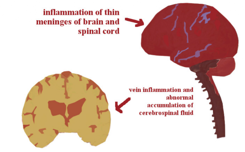
A few of the questions in your mind should at least be: what is most likely to kill this patient, do I need to get a CT of the head, does he need a lumbar puncture, does he need any urgent medications, and what labs should I order?
Meningitis: Clinical Pearls and Pitfalls Read More »
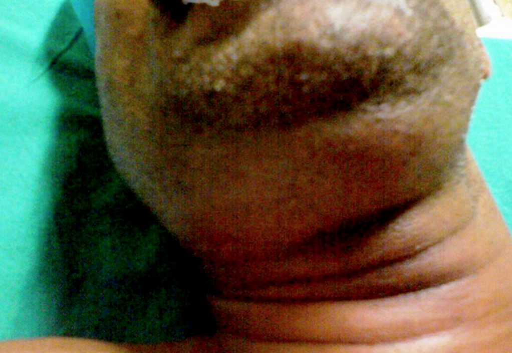
How can you best manage the patient with Ludwig’s Angina?
Ludwig’s Angina: Pearls, Pitfalls, and Highlights Read More »
Featured on #FOAMED REVIEW 32ND EDITION – Thank you to Michael Macias from emCurious for the shout out! Author: Erik A Berg (EM Resident Physician, Keck School of Medicine/Department of Emergency Medicine, LAC+USC Medical Center)//Editors: Jennifer Robertson MD, Alex Koyfman MD BACKGROUND The mosquito-born chikungunya virus (CHIKV) – a cause of an acute onset fever and polyarthralgias – was discovered in sub-Saharan Africa in 1952 and has subsequently been implicated in outbreaks in countries in Africa, Asia, Europe, and the Indian Ocean. As of late 2013, locally transmitted, laboratory confirmed infections have occurred in the Western Hemisphere, including in the United States in July 2014. Of the more than 800,000 suspected and confirmed cases in the Americas, over 80% have occurred in four Caribbean states: Dominican Republic, Martinique, Guadeloupe, and Haiti. The virus is transmitted by mosquito species (Aedes Agyptiae and Aedes Albopictus) that are found throughout the Americas, including in both urban and rural parts of the United States. These same mosquitoes also serve as the primary vector for the dengue virus. CLINICAL The incubation period for CHIKV is typically 2-4 days3, but CHIKV should be suspected in any patient with an acute onset fever, severe polyarthralgias and recent travel to endemic/epidemic areas within 2 weeks of symptom onset. Patients may variably present with nonspecific symptoms including maculopapular rash, headache, myalgias, and conjunctivitis. Shortly after the onset of fever, the majority of infected persons develop severe, often debilitating polyarthralgias. The polyarthralgias are characteristically bilateral and symmetric and most commonly located in small joints (ankle, wrist, hand) and can last for weeks to months. The differential diagnosis for an acute fever with polyarthralgias should include both infectious and rheumatologic conditions. Most importantly for emergency physicians, there are three potentially fatal infectious diseases that can present similarly to CHIKV and share overlapping geographic distributions (1) Leptospirosis: can cause acute fever, jaundice, myalgias localized mainly in calves, and conjunctival suffusion. Conjunctival suffusion and myalgias are considered pathognomonic of leptospirosis. Serology most commonly confirms the clinical diagnosis. (2) Dengue fever: can cause malaise, headache (especially in the retro-orbital area), and muscle aches. The diagnosis is established clinically and confirmed with serology . (3) Malaria: can present with paroxysms of chills and rigor followed by fever spikes, and other nonspecific symptoms including headache, fatigue, myalgia, and nausea. Diagnosis is by direct microscopy (“thick and thin” blood smears). In fact, “chikungunya” is derived from a word in a Tanzanian dialect meaning “that which bends up,” which refers to the bent or stooped posture that infected patients take due to their joint pain. (Ref: Burt FJ, Rolph MS, Rulli NE, et al. Chikungunya: a re-emerging virus. Lancet. 2012;379:662-671) Conjunctival suffusion should be differentiated from conjunctival injection (non-uniform redness) or subconjunctival hemorrhages (Ref: Travel-Acquired Leptospirosis) Laboratory workup for CHIKV is generally non-specific but may show lymphopenia (as in many arboviral diseases). TREATMENT Treatment for CHIKV is limited to supportive care: rest, fluids, antipyretics, and analgesics. DISPOSITION Patients being discharged should be instructed on mosquito control and avoidance. The Aedes mosquitoes are known to be aggressive daytime biters. Patients infected with chikungunya virus should avoid further mosquito exposure during the first week of illness to reduce the risk of further transmission. In addition, patients should be introduced to the possibility of chronic or relapsing polyarthralgic symptoms. References: Centers for Disease Control and Prevention. (2014). Chikungunya, Information for Vector Control. Retrieved from http://www.cdc.gov/chikungunya/pdfs/CHIKV_VectorControl.pdf PAHO (2014). Number of reported cases of Chikungunya fever in the Americas – EW 46. Retrieved from http://www.paho.org/hq/index.php?Itemid=40931 Chen LH, Wilson ME. Dengue and chikungunya infections in travelers. Curr Opin Infect Dis. 2010 Oct;23(5):438-44. Wattal C, Goel N. Infectious disease emergencies in returning travelers: special reference to malaria, dengue fever, and chikungunya. Med Clin North Am. 2012 Nov;96(6):1225-55. Ibid. Morens DM, Fauci AS. Chikungunya at the door–déjà vu all over again? N Engl J Med. 2014 Sep 4;371(10):885-7. doi: 10.1056/NEJMp1408509. Epub 2014 Jul 16. World Health Organization. Guidelines on clinical management of chikungunya fever. Retrieved from http://www.wpro.who.int/mvp/topics/ntd/Clinical_Mgnt_Chikungunya_WHO_SEARO.pdf Centers for Disease Control and Prevention. (2014). Chikungunya, Information for Vector Control. Retrieved from http://www.cdc.gov/chikungunya/pdfs/CHIKV_VectorControl.pdf
D-List Superbugs: Chikungunya Virus Read More »
Often, the dreaded complications that we are taught to look for don’t present to us in the Emergency Department, but develop and evolve during the hospitalization that began with the patient seeing us in the ED. Our skill can help the child in distress, but our vigilance can detect the evolving Acute Chest Syndrome and perhaps even prevent it.
Thanks to Sean M. Fox, MD (@PedEMMorsels) for this gem with significant clinical relevance.
Acute Chest Syndrome Read More »
Questions addressed by EM Lyceum and bulleted by emDocs:
1. When do you use tranexamic acid in trauma?
2. When you can’t get peripheral access in a trauma patient, do you prefer subclavian, femoral, or IO?
3. Which trauma patients do you give PCC to over FFP?
4. In blunt abdominal/flank trauma, do you send a urinalysis or simply look for gross hematuria?
Lyceum Bullets: Trauma Read More »
A discussion of can’t miss life threatening complications in the postpartum period
HELLP!!! Pregnancy Complications in the Postpartum Period Read More »
Stress testing: a beginner’s guide Author: Jason Brown, Capt, USAF, MD (EM Resident Physician, University of Maryland) // Editor: Alex Koyfman, MD Stress tests are aptly named in that the goal is to cause a physiologic stress and to, through a variety of modalities, detect that stress’ impact on the myocardium. There are three major modes of stressing the patient: Exercise – either treadmill or supine bike Vasodilation – adenosine, dipyridamole, regadenoson Inotropy – dobutamine There are five different ways to detect stress on the myocardium: EKG *all modalities employ Echocardiography Radionuclide imaging – Thallium201, Technetium99M PET MRI Treadmill stress testing is the most common form of stress test that you will see as a direct extension of the ED. It employs the Bruce protocol (starting at 1.7mph and 10% grade with increases in both every 3 minutes to a maximum heart rate of 85% (220-age)) while the patient wears an EKG. Tests are positive if the patient has early chest pain, hyper-/hypotension, ST changes, or arrhythmia. Supine bike exercise testing allows for real-time echocardiography. This provides an excellent option for patients with valvular disease and are functional but cannot use a treadmill. The vasodilatory stress tests use agents which increase coronary blood blow. They work on the principle that diseased arteries are already maximally dilated and that there will be no further perfusion of their vascular territories when under stress. A variety of detectors can be used to detect the difference between rest and stress phases. Vasodilation is contraindicated in patients with hypotension, high AV block, or bronchospasm No caffeine (12 hours), Cialis (72 hours), nitrates (48 hours), or calcium channel blockers (48 hours) prior to the test Dobutamine stress tests use the positive inotropic effects of dobutamine to increase the heart rate and elicit perfusion deficits in lieu of actual exercise. There are a variety of protocols but the main goal is to achieve 85% of maximal heart rate (220-age) and to use a detector to examine the myocardium. Contraindicated in patients with arrhythmias, significant hypertension, or LV outflow obstruction. Must hold beta-blockers and calcium channel blockers 24 hours prior. Detection of myocardial perfusion deficits The EKG is the most common modality for detection of ischemic changes. Consistent horizontal or down-sloping ST depressions in contiguous leads is considered positive. In patients that have known CAD or prior revascularization, an abnormal EKG, or a need for functional examination of the heart structures (valvular function, LVEF, etc) then imaging should be considered. There are four major imaging modalities: SPECT, ECHO, PET, MRI. Dobutamine is used in conjunction with echocardiography to evaluate function under stress. New wall motion abnormalities are considered positive for flow-limiting disease. Drawbacks include: technologist-dependent images and difficult interpretation in patients with baseline wall motion abnormalities and/or the obese. All three of the above vasodilators can be used with SPECT, PET, and MRI; deemed myocardial perfusion imaging. All three of these imaging modalities attempt to detect perfusion deficits between rest and stress states. These tests are generally used in patients which need investigations which are beyond the scope of the emergency department. Recommendations: Personally, when I am evaluating a patient in our clinical decision unit (CDU, observation unit) I use either: An EKG treadmill stress for low-intermediate risk ACS patients with normal initial and serial EKGs who can exercise. A supine bike ECHO for any patient that is low-intermediate risk who has an abnormal but nonischemic EKG who can exercise. A dobutamine stress ECHO for any patient with an abnormal EKG who cannot exercise. Any patient with CHF, known CAD, previous PCI/CABG, BBB, or congenital cardiac issue should be evaluated by a staff cardiologist. REFERENCES -Anderson KM, Murphy DL, Balaji M. Essentials of noninvasive cardiac stress testing. J Am Assoc Nurse Pract. 2014;26(2):59-69. -Gibbons RJ, Balady GJ, Bricker JT, et al. ACC/AHA 2002 guideline update for exercise testing: summary article. J Am Coll Cardiol 2002; 40:1531. -Hendel RC, Berman DS, Di Carli MF, et al. ACCF/ASNC/ACR/AHA/ASE/SCCT/SCMR/SNM 2009 Appropriate Use Criteria for Cardiac Radionuclide Imaging. J Am Coll Cardiol 2009; 53:2201. -ACCF/ASE/AHA/ASNC/HFSA/HRS/SCAI/SCCM/SCCT/SCMR 2011 Appropriate Use Criteria for Echocardiography. J Am Soc Echocardiogr 2011; 24:229. -Douglas PS , Khandheria B, Stainback R. et al. ACCF / ASE / ACEP / AHA / ASNC / SCAI / SCCT / SCMR 2008 Appropriateness Criteria for Stress echocardiography. Circulation. 2008;117:1478‐1497 -Fraker TD Jr, Fihn SD, et al. Chronic Stable Angina Writing Committee: focused update of the ACC/AHA 2002 guidelines for the management of patients with chronic stable angina: J Am Coll Cardiol. 2007;50(23):2264. – http://www.ncbi.nlm.nih.gov/pubmed/24730402 – http://www.ncbi.nlm.nih.gov/pubmed/24211281 – http://www.ncbi.nlm.nih.gov/pubmed/23517258 – http://www.ncbi.nlm.nih.gov/pubmed/21908137
Stress testing: a beginner’s guide Read More »