Authors: James Letsen, DO (EM Resident Physician, St. John’s Riverside Hospital Department of Emergency Medicine); Zachary Boivin, MD (Ultrasound Faculty, Yale Department of Emergency Medicine); Jia Jian Li, MD (Ultrasound Faculty, St John’s Riverside Hospital Department of Emergency Medicine) // Reviewed by: Jessica Pelletier, DO, MHPE (APD/Assistant Professor of EM/Attending Physician, University of Missouri-Columbia); Sophia Görgens, MD (EM Physician, Yale University, CT); Cassandra Mackey, MD (Assistant Professor of Emergency Medicine, UMass Chan Medical School); Brit Long, MD (@long_brit)
Welcome to EM@3AM, an emDOCs series designed to foster your working knowledge by providing an expedited review of clinical basics. We’ll keep it short, while you keep that EM brain sharp.
A 95-year-old man with history of aortic stenosis and hyperlipidemia presents after waking with sudden inability to move the fingers and thumb of his right hand. He can lift his arm, flex and extend his wrist normally, and reports intact sensation. He denies numbness, facial droop, dysarthria, headache, gait disturbance, chest pain, or dyspnea.
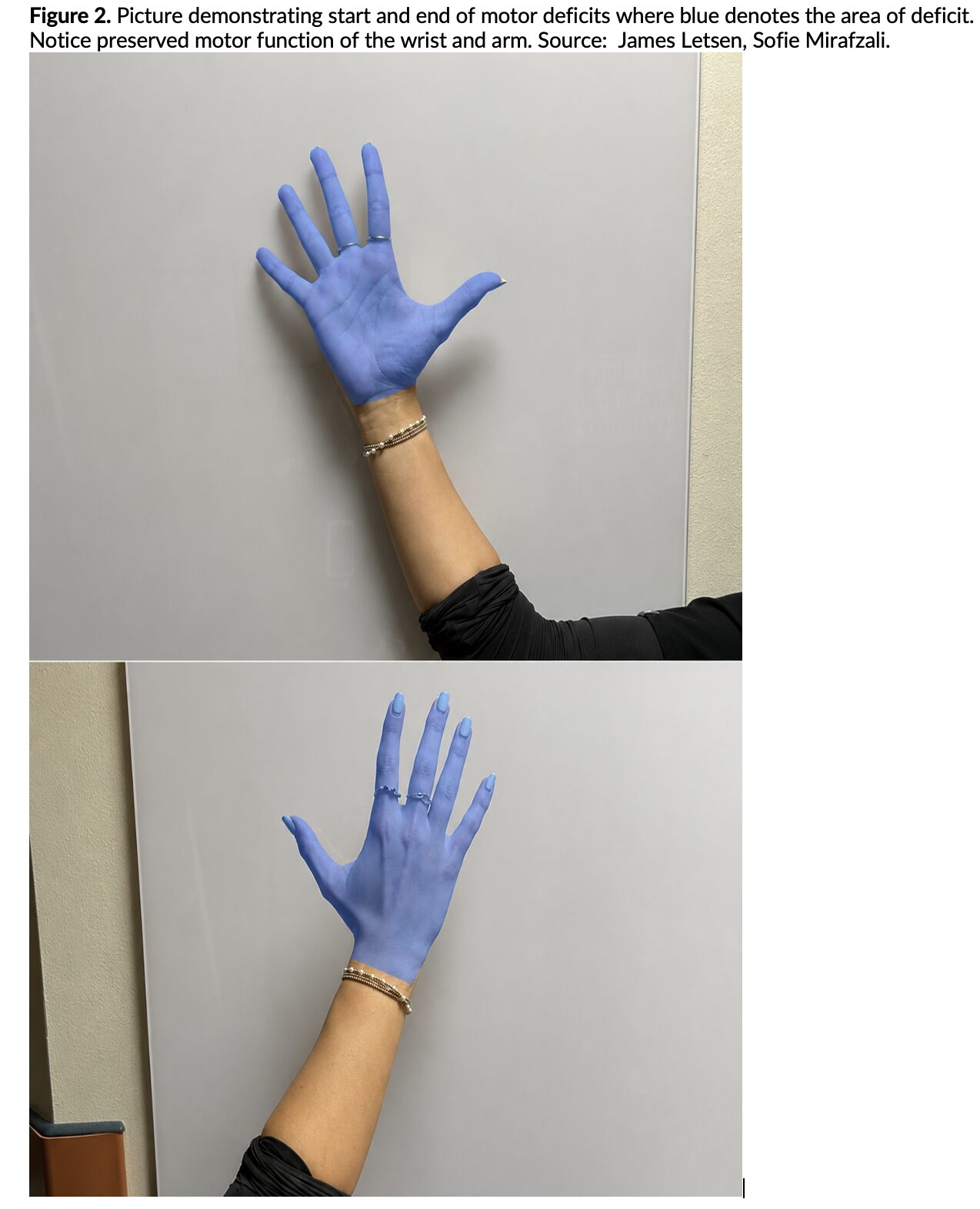
On exam, he has 0/5 strength in finger flexion and extension as well as in the intrinsic hand musculature, with preserved proximal upper extremity strength and intact sensation. He has no other appreciable deficits and normal vital signs. You activate a stroke alert.
Initial non-contrast CT head and CT angiography of the head and neck are unrevealing, and MRI is ordered.
What is the diagnosis?
Answer: Cortical hand-knob stroke mimicking peripheral nerve injury
Background
- The hand-knob region of the precentral gyrus contains the motor representation of the fingers and thumb.
- Small cortical infarcts in this area can produce isolated distal hand paralysis while sparing proximal strength and sensation.
- This presentation closely resembles peripheral nerve injuries and may delay diagnosis if a central process is not considered.1-5
Etiology
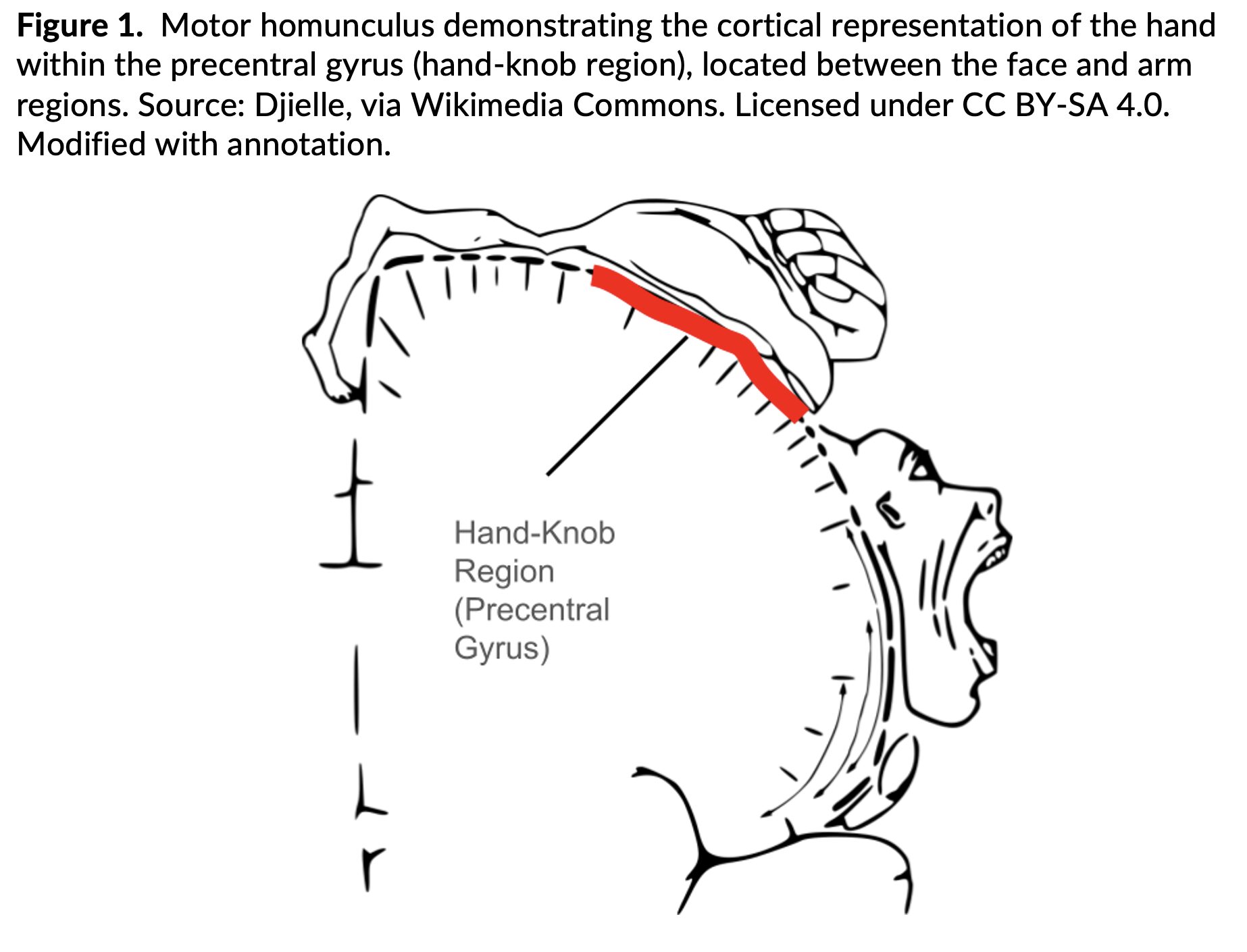
- Hand-knob strokes result from small infarcts involving the precentral gyrus, specifically the region corresponding to hand motor function.
- This area, known as the “hand-knob,” is located along the lateral aspect of the precentral gyrus and represents the motor cortex responsible for fine movements of the fingers and thumb.1-3
- On the motor homunculus, the hand is represented between the face (inferior) and arm (superior) regions. The hand-knob itself has a characteristic omega or epsilon-shaped appearance on axial imaging, which can help localize lesions on MRI.3-6
- These infarcts most commonly arise from:
- Small embolic or thrombotic occlusions of distal cortical branches of the middle cerebral artery
- Cardioembolic disease
- Large-artery atherosclerosis
Small-vessel ischemic disease⁵
- Because this lesion is highly localized to the cortical hand area, patients may present with isolated distal hand weakness without involvement of the face, proximal arm, or sensation, closely mimicking a peripheral nerve injury.1-4,6
Epidemiology
- Hand-knob strokes are uncommon and represent 0.9% of ischemic strokes. 5
- More frequently reported in older adults.
- Commonly associated with vascular risk factors such as hypertension, coronary artery disease, and hyperlipidemia.2-5
Clinical Presentation
- Patients typically present with sudden-onset, painless weakness of the fingers and thumb. Key features include:
- Preserved proximal arm strength (shoulder, elbow) and wrist movement.
- Intact sensation.
- Weakness involving both intrinsic and extrinsic hand muscles.
- No clear dermatomal or single peripheral nerve distribution.1-4,6
- Intrinsic hand muscles (interossei, lumbricals) are responsible for fine motor control within the hand, including finger abduction, adduction, and grip coordination.7,8
- Extrinsic hand muscles originate in the forearm (flexor and extensor compartments) and control finger flexion and extension via long tendons.7,8
- Because the cortical hand-knob region supplies motor input to all of these muscle groups, patients may demonstrate weakness across multiple peripheral nerve territories (median, ulnar, and radial), which is atypical for isolated peripheral nerve injury.1-4,6
- Importantly, palmar sensation is preserved, as sensory pathways are not affected in isolated motor cortex infarcts.1-4 While motor weakness may involve both intrinsic and extrinsic muscles, the absence of sensory deficits helps distinguish this from peripheral neuropathies.
Evaluation and Diagnosis
- Initial non-contrast CT is often normal and primarily serves to exclude intracranial hemorrhage.
- CT angiography is useful to assess for large vessel occlusion, though it is frequently unrevealing in small cortical infarcts.
- For patients presenting within 24 hours of symptom onset, CT perfusion imaging may be considered to evaluate for salvageable penumbra and guide reperfusion decisions. Even isolated hand weakness can represent a functionally disabling deficit,9 particularly in younger or high-functioning patients.
- MRI with diffusion-weighted imaging remains the diagnostic modality of choice and typically demonstrates a small area of restricted diffusion in the precentral gyrus (hand-knob region).1-5
Treatment
- Management follows standard acute ischemic stroke protocols, including rapid stroke team activation, neurologic assessment, and timely imaging to determine eligibility for reperfusion therapy.9
- Patients presenting within 4.5 hours of symptom onset should be evaluated for intravenous thrombolysis, particularly if the deficit is considered functionally disabling. Even isolated hand weakness may qualify if it interferes with activities of daily living or occupational function.9
- For patients outside the thrombolysis window or not meeting criteria, management focuses on secondary stroke prevention, including:
- Antiplatelet therapy
- High-intensity statin therapy
- Blood pressure control
- Further evaluation for a cardioembolic or vascular source is recommended and may include:
- Continuous cardiac monitoring (telemetry) for arrhythmia detection.
- Echocardiography to evaluate for intracardiac thrombus or structural abnormalities.
- Carotid imaging (ultrasound, CTA, or MRA) to assess for atherosclerotic disease
- Prognosis is generally favorable, with many patients demonstrating partial or complete recovery over days to weeks.2-5
Pearls
- Isolated distal hand paralysis with preserved wrist strength and sensation should raise concern for a cortical stroke.
- Peripheral nerve injuries usually follow a single nerve distribution and often include sensory deficits.
- Early CT imaging may be unrevealing; MRI is often required for diagnosis.
- Stroke alert activation for patients presenting <24 hours from symptom onset is appropriate even for isolated motor deficits.
- Early recognition allows timely secondary stroke prevention and improved outcomes.

Which symptom is most consistent with a thrombosis of the anterior cerebral artery?
A) Ataxia
B) Facial droop
C) Homonymous hemianopsia
D) Weakness greater in the lower extremity than the upper extremity
Answer: D
The anterior cerebral artery (ACA) supplies the superior-medial portion of the parietal lobe and the midline portion of the frontal lobe. Occlusion of the ACA accounts for < 3% of all strokes. Symptoms include contralateral motor and sensory loss with weakness greater in the lower extremity than the upper extremity. Facial involvement is not typically seen. Patients can also have confusion and neglect when the dominant hemisphere (typically the left) is affected. Frontal lobe dysfunction can also manifest as altered mentation and impaired judgment.

Ataxia (A) is typically a symptom of posterior circulation occlusion.
A facial droop (B) is seen with involvement of the middle cerebral artery. These patients also have unilateral upper-extremity weakness greater than lower-extremity weakness and sensory loss.
Homonymous hemianopsia (C) (loss of half the visual field on the same side in both eyes) can be seen in middle cerebral artery and posterior cerebral artery strokes.
References
- DeCastro A, Sharma S, Carl RL. Wrist Drop. StatPearls [Internet]. StatPearls Publishing; 2023.
- Chen PL, Hsu TS, Wu YZ, et al. Isolated hand weakness in cortical infarctions. J Formos Med Assoc. 2006;105(8):676-681.
- Alstadhaug KB, Sjulstad A. Isolated hand paresis: a case series. Cerebrovasc Dis Extra. 2013;3(1):65-73.
- Kim JS. Predominant involvement of a particular group of fingers due to small, cortical infarction. Neurology. 2002;58(6):985-989.
- Zhang Z, Sun X, Liu X, Wang L, Zhu R. Clinical features, etiology, and prognosis of hand knob stroke. BMC Neurol. 2022;22:288.
- Takahashi N, Kawamura M, Araki S. Isolated hand palsy due to cortical infarction: localization of the motor hand area. Neurology. 2002;58(9):1412-1414.
- Yousry TA, Schmid UD, Alkadhi H, et al. Localization of the motor hand area to a knob on the precentral gyrus: a new landmark. 1997;120(1):141-157.
- Timsit S, Logak M, Manaï R, et al. Evolving isolated hand palsy: a parietal lobe syndrome associated with carotid artery disease. 1997;120(12):2251-2257.
- Powers WJ, Rabinstein AA, Ackerson T, et al. 2019 Guidelines for the Early Management of Patients With Acute Ischemic Stroke. 2019;50(12):e344–e418