Authors: Michelle Jones, MD (EM Resident Physician, Carolinas Medical Center- Charlotte, NC); Bryant Allen, MD (Associate Program Director/EM Attending Physician, Carolinas Medical Center, Charlotte, NC) // Reviewed by: Sophia Görgens, MD (EM Physician, Yale University, CT); Cassandra Mackey, MD (Assistant Professor of Emergency Medicine, UMass Chan Medical School); Brit Long, MD (@long_brit)
Welcome to EM@3AM, an emDOCs series designed to foster your working knowledge by providing an expedited review of clinical basics. We’ll keep it short, while you keep that EM brain sharp.
A 37-year-old African American female presents to the ED with persistent dry cough and generalized fatigue that has been worsening over the last two months. She has had some mild dyspnea on exertion but denies any fevers or unexplained weight loss. She has had a few episodes that she had attributed to the intensity of her cough.
Initial vital signs include BP of 137/80, HR of 72, RR 18, SpO2 of 95% on RA, T 37.5C. On exam, patient has some diffuse intermittent wheezing and a dry cough. There are a few enlarged lymph nodes in the cervical and axillary regions. She additionally has tender, warm, red, and firm subcutaneous nodules on her bilateral pretibial surfaces.
What is the diagnosis?
Answer: Sarcoidosis
Background:
- Sarcoidosis is a granulomatous syndrome that affects multiple symptoms and has variable presentations.3
- Given the variable presentation of sarcoidosis, it does not have a classic “textbook” description though it is characterized by the formation of granulomas in affected organs.4
Epidemiology:
- First described by English physician Jonathan Hutchinson in 1878 for the cutaneous nodules seen in patients with sarcoidosis and erythema nodosum.5
- Initially called “Mortimer’s Malady”
- Most commonly diagnosed in patients between 35-50 years old.6
- It should be noted that sarcoidosis is rarely diagnosed over the age of 50 and other alternatives should be considered.
- Average incidence rate of 8-11 per 100,000 in the United States.7
- Increased incidence rate in African American patients in the United States at approximately 17-35/ 100,000 people.6
- Some studies have found that sarcoidosis is observed more frequently in women while other studies have shown no difference between men and women.8
- There have been several studies looking at potential environmental and occupational factors that may increase risk for sarcoidosis.
- Many of these studies are limited by misclassification, biases, and confounding factors.9
Clinical Presentation:
- Sarcoidosis presents asymptomatic in approximately 50% of people and can be found incidentally.
- Primarily seen with pulmonary involvement in 90% of cases including dry cough, dyspnea, and chest discomfort.10
- Extrapulmonary presentations of sarcoidosis:
- Cardiac10
- Clinical manifestation 5%
- Cause of death in 20-30% of patients
- Presents with arrhythmias, AV block, dilated cardiomyopathy
- Cutaneous10
- Affecting approximately 20-35% of patients
- Most common – erythema nodosum
- Associated with a more favorable prognosis
- Lupus pernio – unique to sarcoidosis (violaceous, indurated lesions on the face). *Not associated with SLE
- Other presentations including papular, maculopapular, and plaque lesions resembling psoriasis.
- Most common – erythema nodosum
- Affecting approximately 20-35% of patients
- Cardiac10

-
- Ophthalmologic10
- Involved in somewhere between 10 and 60% of cases.
- Uveitis
- Bimodal distribution in the 3rd and 6-7th decades of disease.
- Dry eyes similar to Sjogren’s due to lacrimal sac involvement.
- Uveitis
- Involved in somewhere between 10 and 60% of cases.
- Neurologic10
- Neurosarcoidosis is seen in approximately 5% of patients.
- Primarily cranial nerve dysfunction – specifically the facial nerve causing unilateral facial droop like Bell’s palsy
- Peripheral nerve involvement causing numbness, paresthesias, and weakness
- Leptomeningeal involvement mimicking meningitis with symptoms of ongoing headaches, confusion, and neck stiffness.
- Neurosarcoidosis is seen in approximately 5% of patients.
- Musculoskeletal10
- Löfgren syndrome
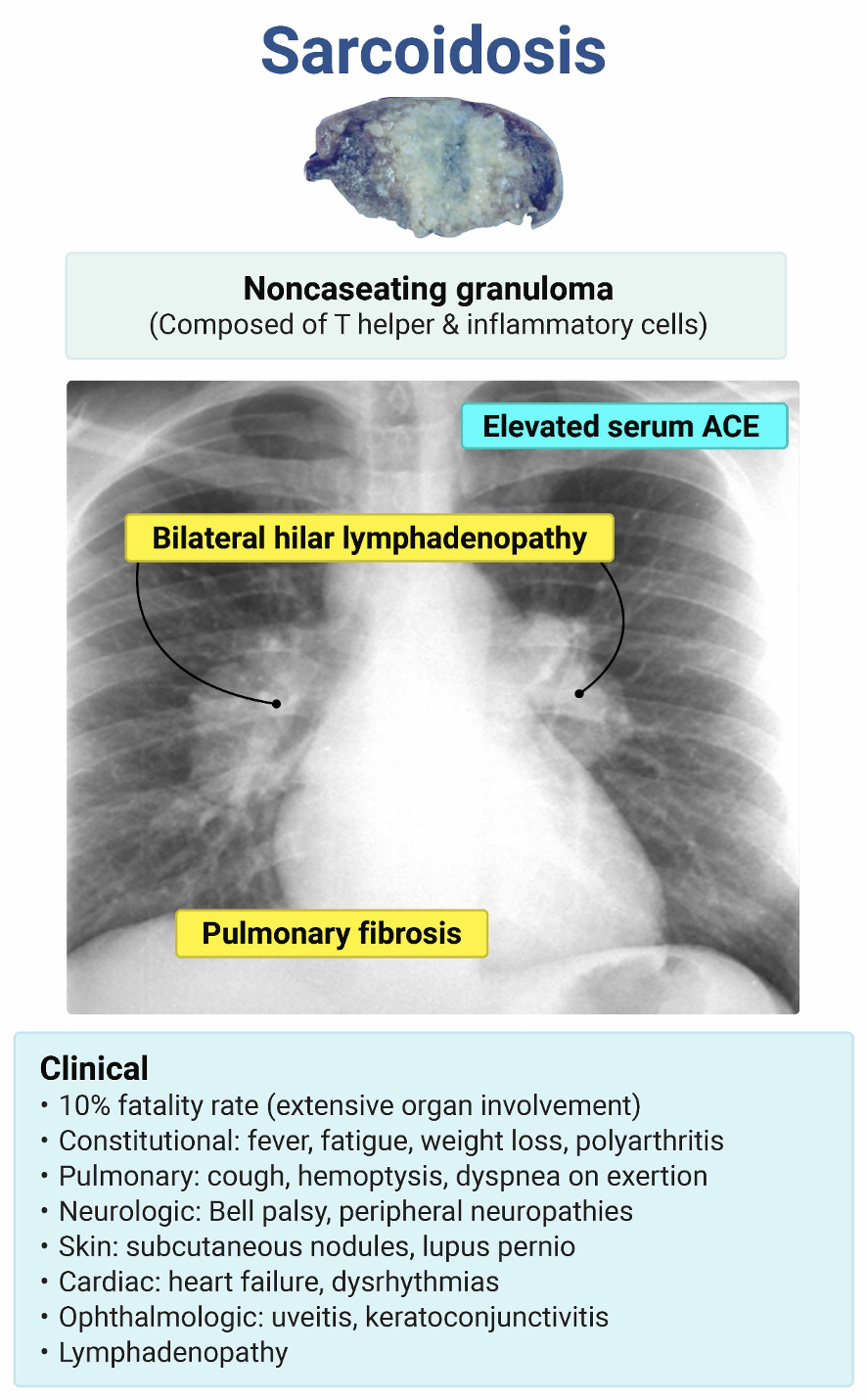
- Bilateral hilar lymphadenopathy, erythema nodosum, fever, and joint involvement.
- Bony lesions occur in approximately 3-13% of cases and sarcoid myopathy in less than 5%.
- Löfgren syndrome
- Gastrointestinal10
- Frequently involving the liver in up to 50-80% of patients.
- Renal10
- Hypercalcemia occurs in 10-20% of sarcoidosis
- Excess conversion of 25(OH) Vitamin D to 1,25(OH) Vitamin D.
- Hypercalcemia occurs in 10-20% of sarcoidosis
- Ophthalmologic10
Evaluation:
- Labs
- CBC, CMP (liver involvement), Calcium, 25-hydroxyvitamin D3, UA, serum angiotensin-converting enzyme (ACE) level6
- Imaging
- Chest Xray6
- Stage 0 – Normal CXR
- Stage I – Mediastinal and hilar adenopathy without pulmonary infiltrates
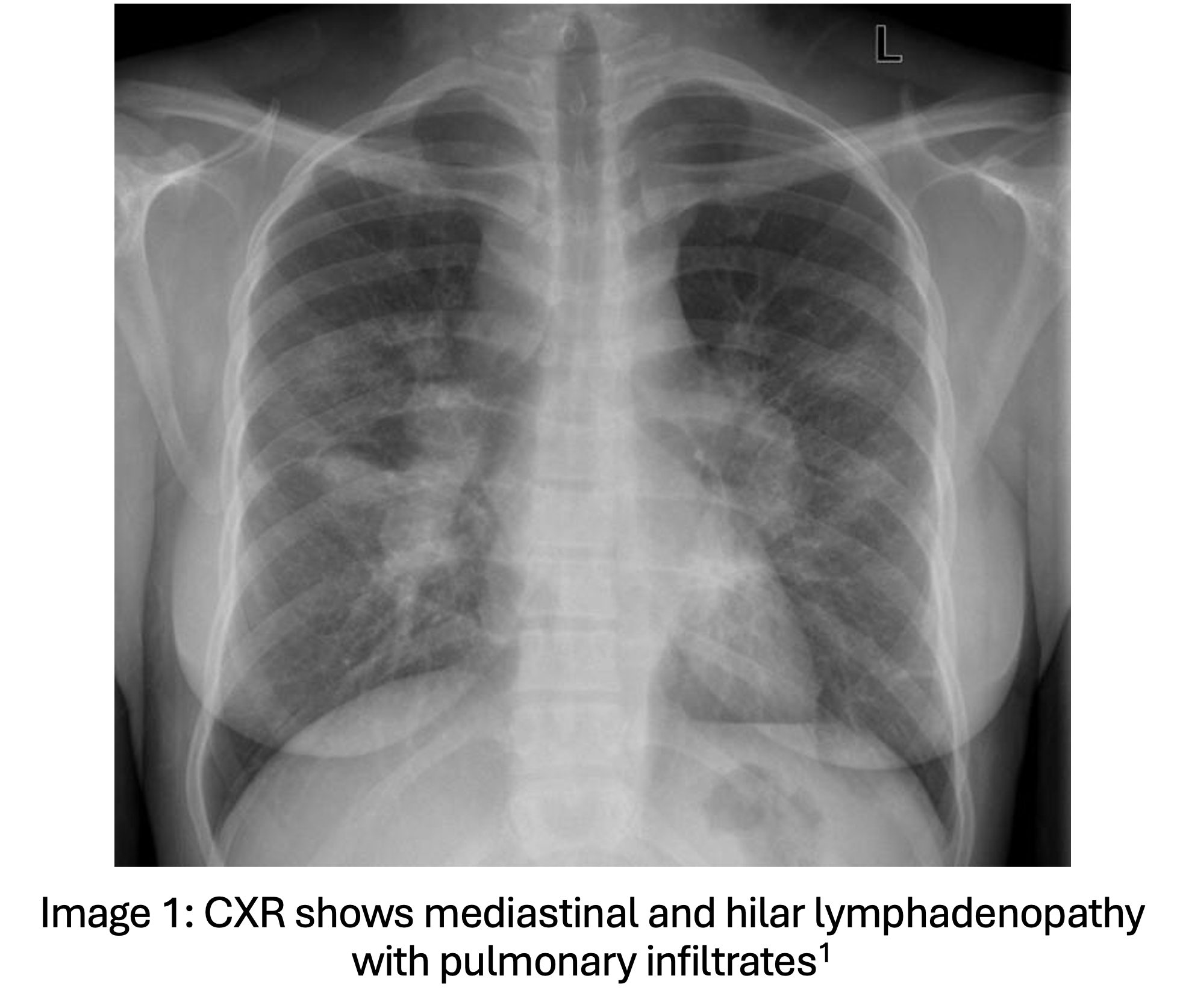
- Stage II – Mediastinal and hilar adenopathy with pulmonary infiltrates
- Stage III – Pulmonary infiltrates without adenopathy
- Stage IV – Pulmonary fibrosis with volume loss, no adenopathy present
- Chest Xray6
- Other tests to consider in the outpatient or inpatient setting6
- Pulmonary function tests (diffusion capacity of the lungs for carbon monoxide (DLCO) and forced vital capacity (FVC))
- Echocardiography, cardiac MRI or PET
- Biopsies
Diagnosis:
- Diagnosis requires all of the following:6
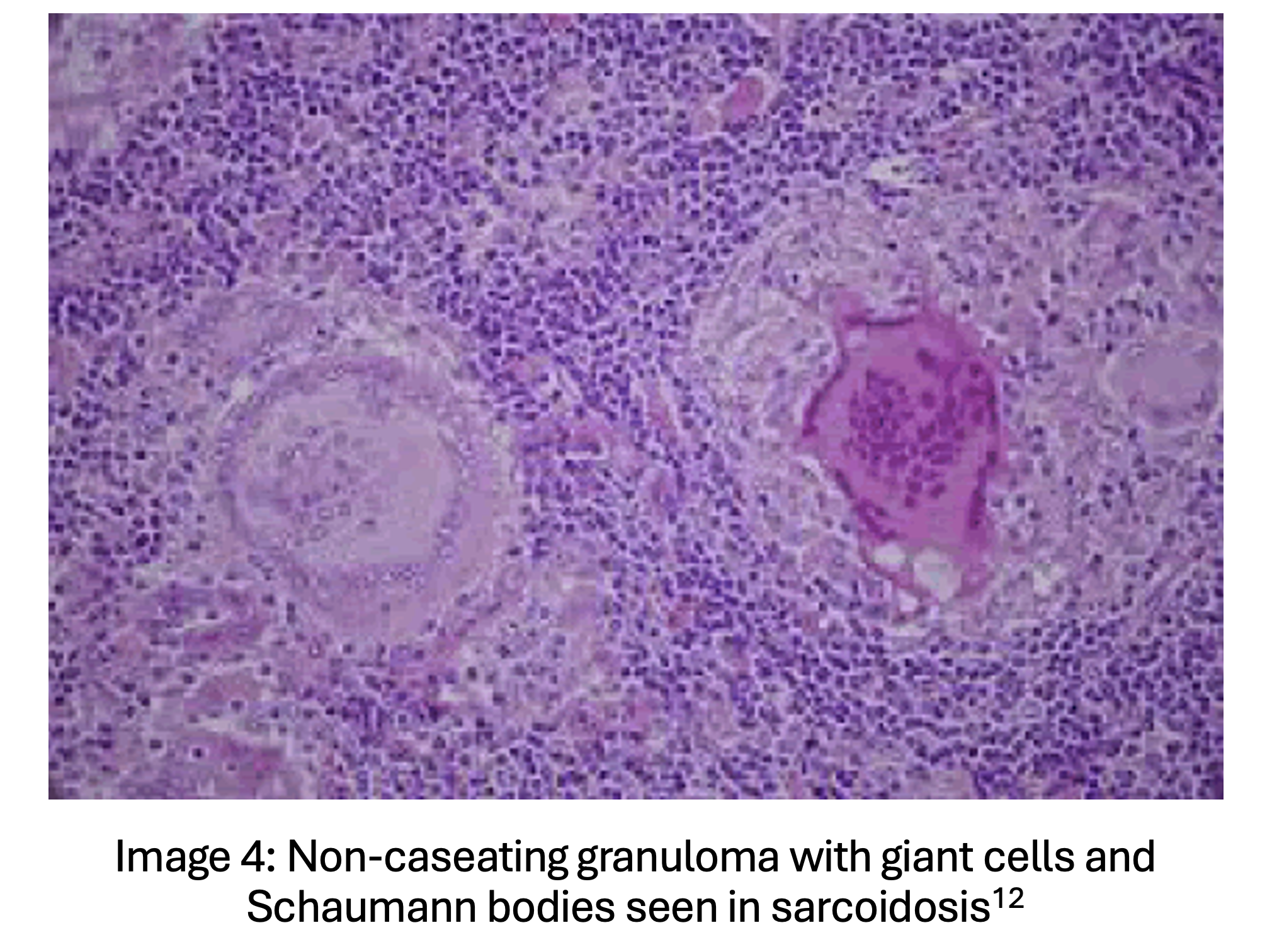
- Noncaseating granulomas on histopathological evaluations.
-
- Clinical symptoms concerning for sarcoidosis.
- Exclusion of other causes of granulomatous disease such as tuberculosis, coccidiomycosis, histoplasmosis, Blastomyces, granulomatosis with polyangiitis, and chronic granulomatous disease.
- Stage I pulmonary sarcoidosis and Löfgren syndrome can be diagnosed without histopathologic evaluation of noncaseating granulomas in the presence of bilateral hilar adenopathy.6
Treatment:
- Treatment for pulmonary sarcoidosis6
- First line: systemic glucocorticoids
- Dosing depends on disease severity – oral prednisolone 20 mg/day in high risk, 5-10 mg/day intermediate.13
- Can consider involving pulmonology or rheumatology for further medications.
- Disease-modifying antirheumatic drugs (DMARDs) – Methotrexate, azathioprine, mycophenolate, and cyclosporin.
- Often preferred for corticosteroid sparing properties.
- Biologic agents are considered a 3rd line option – infliximab, adalimumab.
- Disease-modifying antirheumatic drugs (DMARDs) – Methotrexate, azathioprine, mycophenolate, and cyclosporin.
- First line: systemic glucocorticoids
- Treatment for extrapulmonary sarcoidosis6
- Similar to pulmonary sarcoidosis with additional supportive care specific to each system.
- Ocular – Cycloplegic eye drops
- Joint – NSAIDs
- Similar to pulmonary sarcoidosis with additional supportive care specific to each system.
Prognosis:
- Sarcoidosis often spontaneously remits in nearly half of the cases observed occurring more often in Stage I at a rate of 55-90% and less frequently in Stage II, 40-70%, and Stage III, 10-30%. At Stage IV, irreversible fibrosis is observed and there is no remission observed.14
- Mortality of approximately 1-5% due to complications of the disease however morbidity varies significantly depending on organ involvement and the stage the disease is discovered in.15
Pearls:
- Pulmonary presentations of sarcoidosis are by far the most common, however there are a large variety of extrapulmonary systemic manifestations of sarcoidosis.
- Consult specialist for patients with confirmed or suspected sarcoidosis to monitor disease progression and obtain formal testing to evaluate for extrapulmonary manifestations of sarcoidosis.
- Consider other diagnoses in elderly patients with no prior history of sarcoidosis as sarcoidosis is unlikely to present newly in patients older than 50 years.
- Patients with concerning presentations of sarcoidosis may require admission for further testing and initiation of DMARDs or other biologic medications.

A 45-year-old woman presents to the emergency department for the evaluation of progressive shortness of breath. Over the past 7 months, she has had mild shortness of breath that is made worse with exertion and associated with an occasional dry cough. Her HR is 90 bpm, BP is 142/90 mm Hg, RR is 18/min, and SpO2 on room air is 97%. A chest X-ray is performed and shows bilateral hilar adenopathy with scattered reticulonodular opacities. Which of the following physical examination findings is most likely to be present?
A) Axillary lymphadenopathy
B) Bilateral wheezing and rales
C) Cardiac murmur
D) Digital clubbing
E) Papules on the face and posterior scalp
Answer: E
This patient is presenting with the most common manifestations of sarcoidosis. Sarcoidosis is an inflammatory process that leads to the formation of granulomas within various organs. There are thought to be both genetic and environmental risk factors for sarcoidosis, but no clear pathway leading to the condition has been identified. The most common symptoms of sarcoidosis are chronic and slowly progressive shortness of breath, fatigue, and cough.
Bilateral hilar adenopathy on chest X-ray is the most common finding on evaluation. Cutaneous manifestations are the most commonly detected physical examination finding and one of the most common extrapulmonary manifestations of sarcoidosis. These lesions have a wide range of morphologies and are often chronic. They are most commonly seen on the face, back of the neck, and sites of previous superficial skin trauma. Other less common extrapulmonary findings of sarcoidosis include cardiac dysrhythmia, heart block, right-sided heart failure due to pulmonary hypertension, peripheral lymphadenopathy, and erythema nodosum.
A formal diagnosis of sarcoidosis requires a biopsy to rule out granuloma formation secondary to atypical mycobacterial infection and fungal infection. In patients who are clinically stable without hypoxia, abnormal cardiac rhythms, or rapidly progressive symptoms, this evaluation is appropriate for the outpatient setting.
Axillary lymphadenopathy (A) would not be expected in a patient with sarcoidosis. The most common site of palpable lymphadenopathy in patients with sarcoidosis is the cervical lymph nodes. Cervical lymphadenopathy is another classic sign seen in patients with sarcoidosis.
Bilateral wheezing and rales (B) is an uncommon finding in patients with pulmonary sarcoidosis. While some patients with advanced disease may present with these findings, they are often absent on physical examination.
A cardiac murmur (C) may rarely be present in patients with severe cardiac disease from sarcoidosis. Cardiac sarcoidosis more commonly results in conduction abnormalities and dysrhythmias.
Digital clubbing (D) is seen in the setting of sarcoidosis, primarily in patients with severe pulmonary hypertension. This is a relatively uncommon manifestation of sarcoidosis and, even without treatment, only presents in a small percentage of patients.
Further Reading:
Further FOAMed:
https://emcrit.org/ibcc/sarcoidosis/
Resources:
- Radswiki T, Thoracic sarcoidosis – stage II. Case study, Radiopaedia.org (Accessed on 09 Apr 2026) https://doi.org/10.53347/rID-11908
- Kwong Y, Galaxy sign of sarcoidosis. Case study, Radiopaedia.org (Accessed on 09 Apr 2026) https://doi.org/10.53347/rID-29251
- Llanos O, Hamzeh N. Sarcoidosis.Med Clin North Am. 2019;103(3):527-534. doi:10.1016/j.mcna.2018.12.011
- Culver DA. Sarcoidosis.Immunol Allergy Clin North Am. 2012;32(4):487-511. doi:10.1016/j.iac.2012.08.005
- Young RC Jr, Rachal RE, Cowan CL Jr. Sarcoidosis–the beginning: historical highlights of personalities and their accomplishments during the early years. J Natl Med Assoc. 1984;76(9):887-896.
- Ungprasert P, Ryu JH, Matteson EL. Clinical Manifestations, Diagnosis, and Treatment of Sarcoidosis.Mayo Clin Proc Innov Qual Outcomes. 2019;3(3):358-375. Published 2019 Aug 2. doi:10.1016/j.mayocpiqo.2019.04.006
- Baughman RP, Field S, Costabel U, et al. Sarcoidosis in America. Analysis Based on Health Care Use. Annals of the American Thoracic Society. 2016;13(8):1244–52.
- Brito-Zeron P, Kostov B, Superville D, et al. Geoepidemiological big data approach to sarcoidosis: geographical and ethnic determinants. Clin Exp Rheumatol. 2019;37(6):1052–64.A thorough review of the variation in sarcoidosis clinical characteristics from cohorts around the world
- Arkema EV, Cozier YC. Sarcoidosis epidemiology: recent estimates of incidence, prevalence and risk factors. Curr Opin Pulm Med. 2020;26(5):527-534. doi:10.1097/MCP.0000000000000715
- Rao DA, Dellaripa PF. Extrapulmonary manifestations of sarcoidosis. Rheum Dis Clin North Am. 2013;39(2):277-297. doi:10.1016/j.rdc.2013.02.007
- Lin M. Erythema nodosum SM – aliem. Academic Life in Emergency Medicine. July 16, 2014. Accessed June 1, 2026.
- Bajaj A. Infective germination: Granulomatous inflammation: Lymph node. OMICS International. September 24, 2018. Accessed June 1, 2026.
- Dhooria S, Sehgal IS, Agarwal R, et al. High-dose (40 mg) versus low-dose (20 mg) prednisolone for treating sarcoidosis: a randomised trial (SARCORT trial). Eur Respir J. 2023;62(3):2300198. Published 2023 Sep 9. doi:10.1183/13993003.00198-2023
- Bilgin B, Bilgin MK, Erol S, Celik G, Ozdemir Kumbasar O. Prognosis of sarcoidosis and factors affecting prognosis. Sarcoidosis Vasc Diffuse Lung Dis. 2023;40(4):e2023054. Published 2023 Dec 20. doi:10.36141/svdld.v40i4.13244
- Gerke AK. Morbidity and mortality in sarcoidosis. Curr Opin Pulm Med. 2014;20(5):472-478. doi:10.1097/MCP.0000000000000080