Authors: Adam Engberg, MD (@BlueRidgeEM, EM Resident Physician, Virginia Tech Carilion, Roanoke, VA); Robert Brown, MD (EM Attending Physician, Assistant Professor of Emergency Medicine, Virginia Tech Carilion School of Medicine, Roanoke, VA); and Andrew Moore, MD (@AndrewMooreMD, Assistant Professor of Emergency Medicine, Virginia Tech Carilion School of Medicine, Roanoke, VA) // Reviewed by: Tim Montrief, MD (@EMinMiami); Alex Koyfman, MD (@EMHighAK); Brit Long, MD (@long_brit)
Case 1:
EMS arrives with an obtunded middle-aged male who was complaining of shortness of breath prior to 911 dispatch. His oxygen saturations were in the low 50s upon initial evaluation via EMS; he is saturating 96% on a non-rebreather when he arrives in the emergency department. The decision is made to intubate the patient due to hypoxic respiratory failure and inability to protect his airway. The patient’s O2 saturation declines just prior to successful intubation; it continues to drop despite visual confirmation of ETT passing through the cords, good color change on color capnography, condensation in the tube and confirmation of bilateral breath sounds. The intern’s head is whirling as the patient’s O2 saturation begins to slowly return to physiologically appropriate levels. What should be done next? What is the ED physician’s mental map to correct dropping O2 saturation?
Case 2:
A 37-year-old female presents via EMS after being found unresponsive in the restroom of a local fast food restaurant. The patient was given 2 mg of intranasal naloxone and transported to the local emergency department where she soon became somnolent with decreased respirations. The patient was given an additional 0.4 mg of intravenous naloxone. She was placed on cardiac monitors and plethysmography. The physician and nursing team were called overhead for a new trauma arriving in the department. As the physician team is finishing a long trauma resuscitation, a nurse comes in to let the physician know that the patient who had overdosed on heroin is hypoxic to 87% and apneic. What happened? How could the team have identified an earlier decline in this patient?
Background
Pulse oximetry or SpO2 – where ‘S’ represents saturation and ‘p’ represents pulse – is a mainstay of evaluating oxygenation. Its use is ubiquitous in the Emergency Department (ED), providing a quick, accurate and economical depiction of the level of oxygenated and deoxygenated blood. Indeed, a recent study found that use of SpO2 as part of a routine surveillance program in an orthopedic unit reduced the number of codes, airway alerts, and Intensive Care Unit (ICU) transfers.1
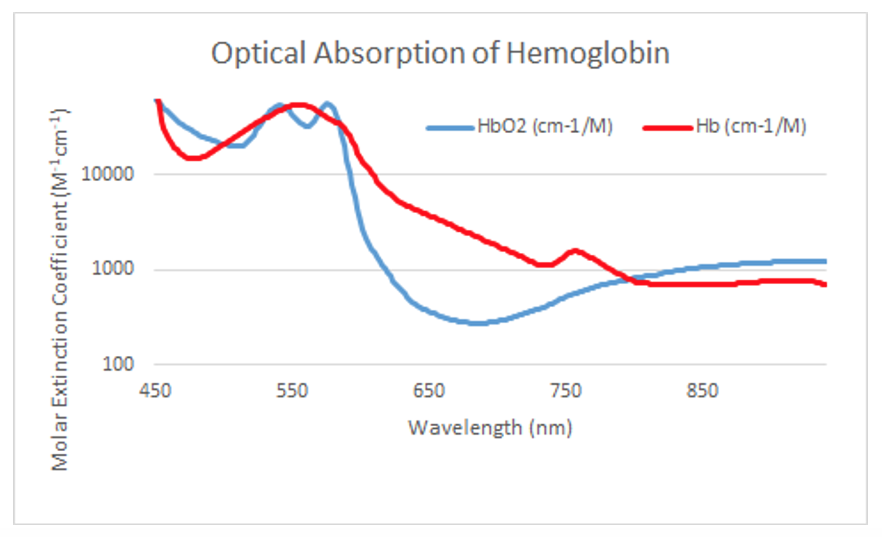
Functionally, the pulse oximeter detects two specific wavelengths that allows differentiation between oxygenated and deoxygenated hemoglobin: Oxygenated blood absorbs more infrared light at 940 nm whereas deoxygenated blood absorbs more 660 nm red light. The wavelength of 660nm is particularly useful because it represents a maximally distinguishable difference between oxygenated and deoxygenated hemoglobin (Figure 1). The pulse oximetry sensor or photodiode cycles between these wavelengths to capture the oxygenation/deoxygenation ratio. An SpO2 of 94% means that 94% of the blood is oxygenated and 6% is deoxygenated.

Figure 1. Optical absorption of hemoglobin. Figure recreated with permission from publicly available data compiled by Dr. Scott Prahl, Oregon Medical Laser Center: https://omlc.org/spectra/hemoglobin/summary.html
Limitations and Pitfalls
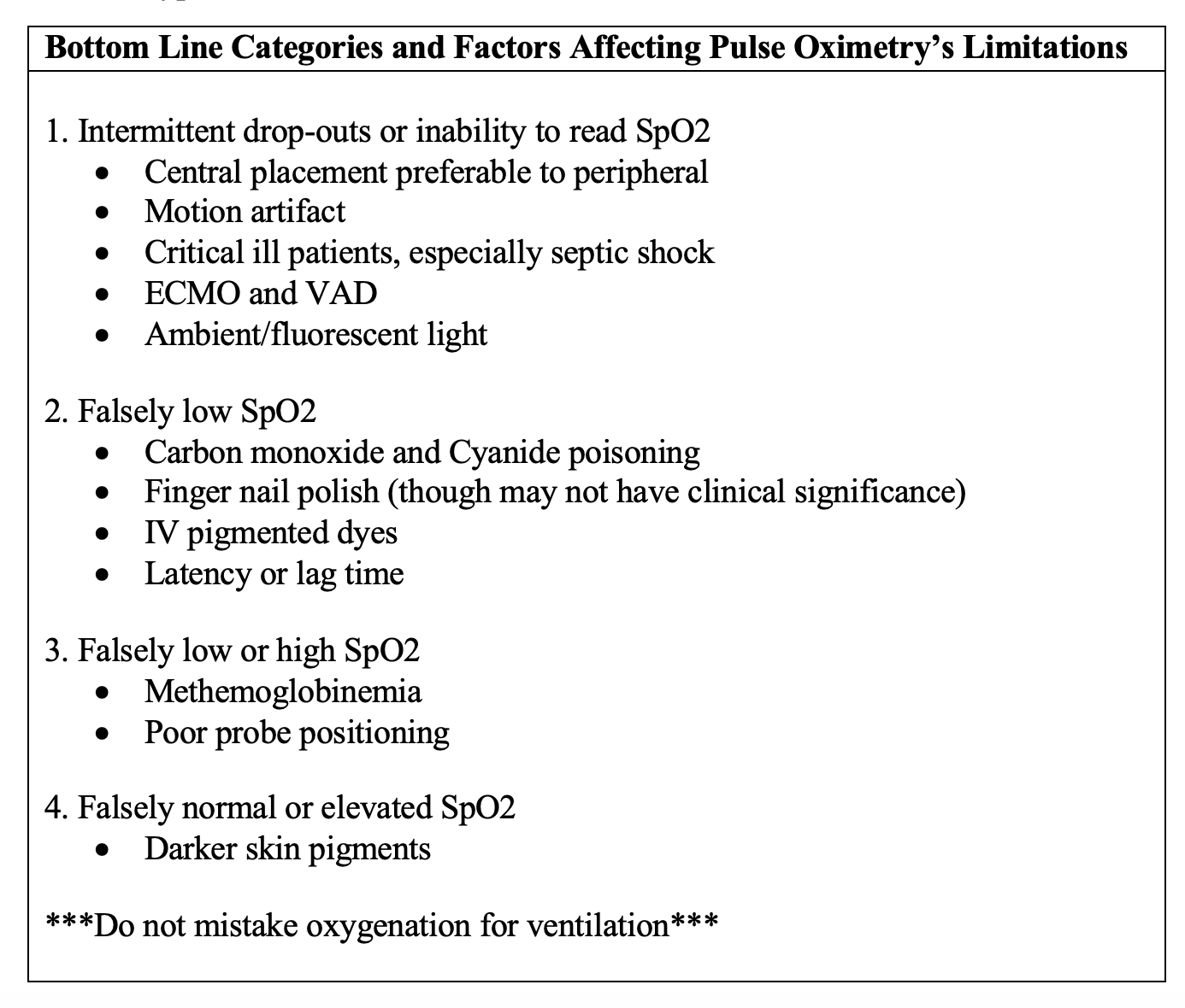
The SpO2 can be misleading. Even in healthy patients, it lags behind the clinical onset of hypoxia by more than a minute,2 and the delay may be longer with finger probes than ear probes3 or forehead probes.2 Limited data indicate a centrally placed pulse oximeter performs better than a peripheral one, but neither is capable of hypopneic hypoventilation detection.4 In one small study, end-tidal CO2 decreased almost 4 minutes before decreases in SpO2 on average.5
Falsely reassuring SpO2 is obviously dangerous and two important examples arise from exposure to fires: carbon monoxide and cyanide poisoning. Carbon monoxide forms carboxyhemoglobin which mimics the absorption frequency of oxyhemoglobin, creating the appearance of well-oxygenated blood despite hypoxia.6 Co-oximetry detects carboxyhemoglobin and pulse co-oximetry shortens the time to detection and treatment in the ED,7 though it serves best as a screening tool rather than a substitute for serum carboxyhemoglobin tests.8 Cyanide decouples oxidative phosphorylation so the patient is dependent on anaerobic metabolism despite normal oxygenation, leading to profound acidosis and death.9 While cyanide does not interfere with SpO2, hydroxocobalamin, the bright red antidote interferes with co-oximetry and makes subsequent measurements of carboxyhemoglobin unreliable.10
As the hydroxocobalamin and carboxyhemoglobin (COHb) examples demonstrate, conditions which alter the color of serum (injecting dyes) or the color of hemoglobin (anemias and hemoglobinopathies) alter the SpO2. The degree of SpO2 depression and the duration of the effect after giving a dye may be more severe in cases of concomitant anemia.11Regardless of hemoglobin level or reticulocyte count, sickle cell vaso-occlusive crisis falsely decreased the SpO2 roughly 4% in one study,12 as seen with certain rare genetic variant hemoglobins as well.13 Methemoglobinemia can prompt both unnecessary intervention and spurious pulse oximetry readings. Even with an elevated partial pressure of oxygen (PaO2), it reduces SpO2 to ~85%.14 When the PaO2 does decrease, the SpO2 fails to decrease commensurately, giving false reassurance.15 In one example: ‘at COHb = 70%, the SpO2 is approximately 90% when inspired oxygen fraction (FIO2) is 1.0, whereas the actual SaO2 is only 30%.16 Not all pigments and hemoglobin variants depress SpO2. Neither bilirubin17 nor fetal hemoglobin16 appears to have an impact as once believed.
Because the oximeter relies on pulsatility to differentiate arterial blood for analysis, conditions which generate venous pulsatility or which diminish arterial pulsatility make SpO2 unreliable. Of all the conditions with an unpredictable impact on SpO2, septic shock is the most important to recognize. A retrospective study of 88 consecutive subjects found an overestimation of pulse oximetry by 2.75% in patients with severe sepsis and septic shock.18 Continuous flow left ventricular assist devices (LVADs) operate at speeds which are unable to garner sufficient pulsatility, requiring arterial blood gas (ABG) measurements or cerebral oximetry as alternatives.19,20 Poor waveforms can also adversely affect accuracy in patients on Extracorporeal Membrane Oxygenation (ECMO).21 Pulse oximetry can overestimate oxygen saturation in patients on veno-venous ECMO (VV-ECMO) due to rising COHb levels.22 Motion of the hand similarly undermines the ability of the probe to detect pulsatility accurately, resulting in increased SpO2 variability.23
Outside the blood vessels, a number of environmental factors affect SpO2, but are less likely to be clinically significant. Some light sources in the room produce wavelengths measured by the pulse oximeter, but none alters the SpO2 by more than 5%.24 Fingernail polish can depress the SpO2 without a clinically important impact on average,25 but with a wide variance, greatest for dark shades like black, blue, and purple.26 Rotating the probe 90 degrees – such that the diode emanates through the sides of the fingers – can serve as a remedy should placement over the fingernail prove troublesome.27
Pulse oximetry’s shortcoming in providing accurate readings in darker skinned individuals is particularly alarming. Two recent large cohorts found the correlation of pulse oximetry and arterial blood gases of Black patients revealed almost three times the frequency of occult hypoxemia.28 Indeed, the use of pulse oximetry to triage patients of color may result in missed cases of hypoxemia.

Case Conclusions
Case 1: The O2 levels rebounded to a normal level and the patient’s O2 requirement on the vent was slowly weaned with serial ABG checks. The patient was ultimately admitted to the ICU after intubation and initiation of broad-spectrum antibiotics for presumed drug resistant pneumonia. The SpO2 takes time to register hypoxemia, even in healthy volunteers. This delay is more significant in critical patients with hypoperfusion and peripheral vasoconstriction.
Case 2: The patient was ventilated with a bag valve mask and 400 mcg of naloxone was injected via IV. The patient became responsive again, demonstrating an ability to breath on her own. She revealed using methadone today in addition to recent heroin use. She was started on a naloxone infusion at 250 mcg per hour and admitted to the step-down unit for further management of opiate overdose and airway monitoring. She was placed on end-tidal capnography.
The patient likely became more and more apneic due to the short half-life of naloxone. Because pulse oximetry does not monitor ventilation, there will be a delay in identifying patients who become hypoxic secondary to apnea. Early use of end-tidal CO2 is paramount when attempting to monitor ventilation.
Summary
Pulse oximetry is a standard in monitoring the vital signs of emergency department patients around the world. It is imperative to understand how these sensors work. More importantly, though, is anticipating how and when pulse oximetry may mislead us. The cases we chose highlight two significant flaws in peripheral pulse oximetry: latency and ventilation.
Further Reading:
FOAMed:
Weingart, S. EMCrit Podcast 88 – Oxygen Physiology with Daniel Davis. December 10, 2012. https://emcrit.org/emcrit/oxygen-physiology/. Accessed August 30, 2020.
Orman, R. How to Use the Pulse Ox Like a Boss. From Essentials of Emergency Medicine NYC 2017, Strayer, R. March 11, 2019. https://ercast.libsyn.com/how-to-use-the-pulse-ox-like-a-boss. Accessed August 29, 2020.
References:
- Taenzer AH, Pyke JB, McGrath SP, Blike GT. Impact of pulse oximetry surveillance on rescue events and intensive care unit transfers: a before-and-after concurrence study. Anesthesiology 2010;112(2):282–7.
- Choi SJ, Ahn HJ, Yang MK, et al. Comparison of desaturation and resaturation response times between transmission and reflectance pulse oximeters. Acta Anaesthesiol Scand 2010;54(2):212–7.
- Young D, Jewkes C, Spittal M, Blogg C, Weissman J, Gradwell D. Response time of pulse oximeters assessed using acute decompression. Anesth Analg 1992;74(2):189–95.
- Lindholm P, Blogg SL, Gennser M. Pulse oximetry to detect hypoxemia during apnea: comparison of finger and ear probes. Aviat Space Environ Med 2007;78(8):770–3.
- Langhan ML, Chen L, Marshall C, Santucci KA. Detection of hypoventilation by capnography and its association with hypoxia in children undergoing sedation with ketamine. Pediatr Emerg Care 2011;27(5):394–7.
- Rg B, Se A, Jl E, R R, J S, P S. The pulse oximetry gap in carbon monoxide intoxication [Internet]. Annals of emergency medicine. 1994 [cited 2021 Mar 2];24(2). Available from: https://pubmed.ncbi.nlm.nih.gov/8037391/
- Hampson NB. Noninvasive pulse CO-oximetry expedites evaluation and management of patients with carbon monoxide poisoning. Am J Emerg Med 2012;30(9):2021–4.
- Sebbane M, Claret P-G, Mercier G, et al. Emergency department management of suspected carbon monoxide poisoning: role of pulse CO-oximetry. Respir Care 2013;58(10):1614–20.
- Hall AH, Rumack BH. Clinical toxicology of cyanide. Ann Emerg Med 1986;15(9):1067–74.
- Lee J, Mukai D, Kreuter K, Mahon S, Tromberg B, Brenner M. Potential interference by hydroxocobalamin on cooximetry hemoglobin measurements during cyanide and smoke inhalation treatments. Ann Emerg Med 2007;49(6):802–5.
- Saito S, Fukura H, Shimada H, Fujita T. Prolonged interference of blue dye “patent blue” with pulse oximetry readings. Acta Anaesthesiol Scand 1995;39(2):268–9.
- Comber JT, Lopez BL. Evaluation of pulse oximetry in sickle cell anemia patients presenting to the emergency department in acute vasoocclusive crisis. Am J Emerg Med 1996;14(1):16–8.
- Robertson A, Rahemtulla A. Pulse oximetry error in a patient with a Santa Ana haemoglobinopathy. BMJ Case Rep 2016;2016:bcr2016216787.
- Barker SJ, Tremper KK, Hyatt J. Effects of methemoglobinemia on pulse oximetry and mixed venous oximetry. Anesthesiology 1989;70(1):112–7.
- Feiner JR, Rollins MD, Sall JW, Eilers H, Au P, Bickler PE. Accuracy of carboxyhemoglobin detection by pulse CO-oximetry during hypoxemia. Anesth Analg 2013;117(4):847–58.
- Lebecque P, Shango P, Stijns M, Vliers A, Coates AL. Pulse oximetry versus measured arterial oxygen saturation: a comparison of the Nellcor N100 and the Biox III. Pediatr Pulmonol 1991;10(2):132–5.
- Abrams GA, Sanders MK, Fallon MB. Utility of pulse oximetry in the detection of arterial hypoxemia in liver transplant candidates. Liver Transpl 2002;8(4):391–6.
- Wilson BJ, Cowan HJ, Lord JA, Zuege DJ, Zygun DA. The accuracy of pulse oximetry in emergency department patients with severe sepsis and septic shock: a retrospective cohort study. BMC Emerg Med 2010;10:9.
- Hessel EA. Management of patients with implanted ventricular assist devices for noncardiac surgery: a clinical review. Semin Cardiothorac Vasc Anesth 2014;18(1):57–70.
- Aldrich TK, Gupta P, Stoy SP, Carlese A, Goldstein DJ. Pulseless Oximetry: A Preliminary Evaluation. Chest 2015;148(6):1484–8.
- Farkas J. Top 10 reasons pulse oximetry beats ABG for assessing oxygenation. [Internet]. Top 10 reasons pulse oximetry beats ABG for assessing oxygenation. 2016 [cited 2020 Aug 29];Available from: https://emcrit.org/pulmcrit/pulse-oximetry
- Nisar S, Gibson CD, Sokolovic M, Shah NS. Pulse Oximetry Is Unreliable in Patients on Veno-Venous Extracorporeal Membrane Oxygenation Caused by Unrecognized Carboxyhemoglobinemia. ASAIO J 2020;66(10):1105–9.
- Gehring H, Hornberger C, Matz H, Konecny E, Schmucker P. The effects of motion artifact and low perfusion on the performance of a new generation of pulse oximeters in volunteers undergoing hypoxemia. Respir Care 2002;47(1):48–60.
- Fluck RR, Schroeder C, Frani G, Kropf B, Engbretson B. Does ambient light affect the accuracy of pulse oximetry? Respir Care 2003;48(7):677–80.
- Yamamoto LG, Yamamoto JA, Yamamoto JB, Yamamoto BE, Yamamoto PP. Nail Polish Does Not Significantly Affect Pulse Oximetry Measurements in Mildly Hypoxic Subjects. RESPIRATORY CARE 2008;53(11):5.
- Hinkelbein J, Genzwuerker HV, Sogl R, Fiedler F. Effect of nail polish on oxygen saturation determined by pulse oximetry in critically ill patients. Resuscitation 2007;72(1):82–91.
- Chan ED, Chan MM, Chan MM. Pulse oximetry: Understanding its basic principles facilitates appreciation of its limitations. Respiratory Medicine 2013;107(6):789–99.
- Sjoding MW, Dickson RP, Iwashyna TJ, Gay SE, Valley TS. Racial Bias in Pulse Oximetry Measurement. New England Journal of Medicine 2020;383(25):2477–8.


