
Originally published at EM Cases – Visit to listen to accompanying podcast. Reposted with permission.
Follow Dr. Anton Helman on twitter @EMCases
There are a whole slew of very important occult knee injuries – those that have a normal or near normal x-ray – that can cause serious morbidity if you miss them, and for the catchall soft tissue injuries there are some subtleties in diagnosis and management that will make a real difference to our patients. Arun Sayal and Hossein Mehdian answer questions such as: When should we suspect a spontaneously reduced knee dislocation? Do all patients suspected of a spontaneous knee dislocation require a CT angiogram to rule out vascular injury? Which patients with a low energy mechanism are at risk for knee dislocation and vascular complications? How can you increase the accuracy of the active straight leg raise in assessing for quadriceps and patella tendon rupture? What is an easy way to identify patella baja and patella alta? What are the indications for ultrasound of the knee? What are the true indications for a knee immobilizer and how can knee immobilizers kill our patients? and many more…
General Principles of Occult Knee Injuries
A) A normal X-ray does not rule out a serious knee injury.
There are several important diagnoses that you should consider in a patient who comes in with knee pain and has a normal x-ray of the knee.
- Quads/patellar tendon rupture
- Lateral tibial plateau fracture
- Dislocation w reduction
- Locked knee
- Hip injury with referred pain
- Compartment syndrome
- Septic knee
B) Mechanism of Injury: Direct blow vs valgus strain vs sudden deceleration vs twisting
Direct blow – patella fracture, knee dislocation, tibia fracture
Valgus strain – MCL tear and/or lateral tibial plateau fracture
Sudden deceleration – ACL tear
Twisting – Meniscus tear
C) Age – The same mechanism in patients of different ages can lead to different injuries (e.g., with a valgus force injury to the knee you should consider a distal femur fracture in a child, an MCL injury in a young adult and lateral tibial plateau fracture in an older person)
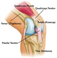
Physical exam pearls for occult knee injuries
Many patients with acute knee injuries in the ED will experience a lot of pain and often clinicians short-change their examination of the knee because they don’t want to cause more pain. However, there are several tips to help the patient relax enough to do provocative testing and the essential maneuvers:
Patient must be supine on a stretcher (not sitting in a chair) with both knees fully exposed.
Place a pillow or roll under the distal femur so that the knee is relaxed at about 20 degrees of flexion to allow for provocative knee testing.
Always perform an active straight leg raise to assess for extensor mechanism function (quadriceps or patella tendon rupture).
“The lateral joint line is the dark corner of the knee exam” – examine the lateral joint line in addition to the medial joint line to assess for the possibility of a lateral tibial plateau fracture in the setting of a valgus mechanism of injury.
Occult Knee Dislocation
About 20-50% of all knee dislocations spontaneously reduce before the patient arrives at the ED.
A significant minority of occult knee dislocations will have neurovascular injuries which if missed, may lead to ischemic complications and even limb amputation.
When to suspect an occult knee dislocation:
- Three of the 4 knee ligament disruption or ligamentous laxity in multiple directions (a “loose knee”)
- A ‘buckling’ knee with a foot drop (common peroneal nerve palsy)
- If upon lifting up the patient’s leg by their heels the knee falls into hyper-extension compared to the contralateral knee
- High energy mechanism of injury or low energy mechanism in patients with BMI>40
Clinical Pearl: Three out of four knee ligament laxity (ACL, PCL, MCL, LCL) is suspicious for an occult knee dislocation
The presence of normal distal pulses and a normal Ankle Brachial Index >0.9 (ABI) does not completely rule out occult popliteal artery injury. If you suspect an occult knee dislocation, refer immediately to orthopedics as revascularization within 6-8 hours of popliteal artery injury is recommended to prevent ischemic complications. While almost all patients with suspected knee dislocation used to get a CT angiogram to assess for vascular injury, the current thinking is that the vast majority of patients who do have a vascular injury with normal serial neurovascular exams and normal serial ABIs have intimal tears that do not require surgery and so these patients can be simply anticoagulated and admitted for observation without a CT angiogram.
Caution: Low-energy trauma can cause knee dislocations, especially in patients with a BMI>40. Obese patients with low-energy trauma were more likely to have associated neurovascular injury than high-energy trauma patients in one study.
Occult Knee Dislocation Take Home
Don’t send home patients who tell you that it felt like their knee popped out of place and they have a big swollen knee with laxity in multiple directions on exam, until you’re sure they haven’t had a vascular injury related to a self-reduced knee dislocation. Even if they have palpable peripheral pulses and a normal ABI, speak to your orthopedic surgeon, consider getting a CT angiogram to rule out a popliteal injury, admit, and consider the same in any obese person even with a low force mechanism.
Reduction of Knee Dislocation
Tibial Plateau Fracture
For patients with a valgus type injury like a pedestrian getting bumped in the side of the knee by a car or a soccer player getting hit in the lateral knee who you’re thinking MCL but they have lateral pain, think tibial plateau fracture and examine the lateral joint line carefully. Occult tibial plateau fractures are not uncommon in older people with a low energy valgus force with axial loading.
“The lateral joint line is the dark corner of the knee exam” – don’t forget to examine the lateral joint line!
Pearl: When assessing for a suspected MCL injury after a valgus force injury and you’re applying valgus strain to the knee, if the patient complains of both lateral and medial knee pain, look for a lateral tibial plateau fracture.

Lateral tibial plateau fracture. Note that the subchondral line is lower on the lateral vs medial side.
X-ray pearls for subtle lateral plateau fractures
- Normally the lateral subchondral line is higher (more proximal) than the medial one; if, on the AP or lateral x-ray, the lateral subchondral line is at a lower horizontal level compared to the medial one, assume a depressed lateral tibial plateau fracture.
- Scrutinize the oblique view for a depressed lateral tibial plateau.
Even if the x-ray doesn’t show that subtle depression of the tibial plateau, the patient should be protective weight bearing: a walker with touch weight bearing in the older person or a knee immobilizer in a younger person and very close ortho follow up, or, if you have the option, go straight to CT. And if you see a depressed tibial plateau fracture of more than a 2mm or a split displaced fracture refer directly to ortho in the ED, as these patients will usually require urgent surgery.
Extensor Mechanism Injuries, The Straight Leg Raise and The Real Indications for Knee Ultrasound and Knee Immobilizers
Quadriceps tendon ruptures are more commonly seen in patients older than 40 years of age while the less frequent patella tendon ruptures are more commonly seen in patients under the age of 40.
Quadriceps tendon rupture is often misdiagnosed as a simple “knee sprain” and missed in up to 50% of patients. It is important to pick these injuries up in the ED because surgery is required within a week or two to prevent retraction of the muscle.
The triad of 1) acute knee pain with difficulty weight bearing, 2) inability to actively straight leg raise and 3) a suprapatellar gap or infrapatellar gap suggest the diagnoses of quadriceps or patella tendon rupture respectively.

Suprapatellar gap of quadraceps tendon rupture
Pearl: To increase the accuracy of the straight leg raise to screen for extensor mechanism injuries such as quadriceps or patellar tendon rupture, ensure that the patient is not externally rotating their leg thereby using their hip abductors to raise their leg injuries, and eliminate the iliotibial band from the leg raise by having the patient sit on the edge of the bed with the lower legs hanging over the edge and ask the patient to straight leg raise.
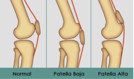
Often patients with quadraceps or patella tendon rupture will have normal x-rays. With a quadriceps tendon rupture, you may see Patella Baja (patella rides lower than usual). In a patellar tendon rupture, you may see Patella Alta (patella rides higher than usual – think alta for high altitude).
Pearl: An easy way to determine patella baja or patella alta is that the patella is normally one finger breadth above the joint line and any deviation from this may indicate a patella baja or alta. The Insall-Salvati ratio is a more exact method to determine patella baja and alta
The only indication for a formal ultrasound for knee injuries is for assessment of the extensor mechanism, not for suspected ![]() meniscus or ligament injuries.
meniscus or ligament injuries.
Indications for knee immobilizer (e.g. Zimmer splint) in soft tissue injuries of the knee
The indications for a knee immobilizer are for extensor mechanism injuries
- First time patella dislocation
- Quadriceps tendon rupture
- Patella tendon rupture
These patients should be placed into a knee immobilizer and followed up with orthopedics because a complete tear usually requires surgery within one week to prevent retraction of the quadriceps.
Knee immobilizers are not indicated for suspected meniscus or ligament injuries of the knee as they do not aid in healing, may increase the rate of falls in older patients and cause atrophy of the quadraceps as well as stiffness leading to decreased range of motion.
“The knee immobilizer in the ED is akin to Amoxicillin in the walk-in clinic”
– Arun Sayal
Occult Knee Injuries: ACL Tear
The diagnosis of ACL tear is usually made on history alone, while the physical exam in the ED is usually non-contributory as the patient often can’t tolerate provocative testing. A good knee exam should be performed nonetheless to assess for the possibility of occult knee dislocation and other injuries.
History: Most ACL tears occur in the setting of sports without contact of another player and the the patient usually needs to be carried off the court or field. The ACL tears when their is sudden deceleration, the knee twists with the tibia pushed anteriorly usually with concurrent valgus stress. 70% of patients report a ‘pop’ and some report buckling or “giving out”. Significant swelling usually occurs within hours (as apposed to meniscus injury which is usually delayed).
Physical: Practically speaking the Lachman test is the only useful physical exam maneuver for assessment of ACL as the anterior drawer and pivot shift cause too much pain in the acute phase for that patient to be able to tolerate them.
What to look for on x-ray in a patient with suspected ACL tear
In pediatric patients with suspected ACL tear, look for tibial spine fracture on both AP and lateral x-ray which will change management in that these patient require immobilization in extension.

Tibial spine fracture seen on lateral view. Case courtesy of Dr Jeremy Jones, Radiopaedia.org. From the case rID: 27372
In adult patients with suspected ACL tear, look for a Segond fracture at the lateral tibial which will confirm the diagnosis but not change management.

Segond fracture confirms the diagnosis of ACL rupture. Case courtesy of Gerry Gardner , Radiopaedia.org. From the case rID: 13910
Management of suspected ACL tear
Avoid knee immobilizers!
Weight bearing as tolerated with early ROM exercises.
Trick of the trade: cut out a circle the size of the patient’s patella from an ABD pad and place the remaining ABD pad over the patient’s knee with a tensor bandage to hold it in place for improved compression of the knee
The R in RICE stands for Restricted Activity, not Rest
For ACL, MCL and meniscus injuries instruct the patient to let pain be their guide, weight bear as tolerated with early ROM exercises to avoid knee stiffness and delay to normal activity.
How to differentiate a spontaneously reduced patella dislocation from an ACL injury
Patients with a spontaneously reduced patella dislocation will often have a history of anterior knee pain.
Usually the medial patello-femoral ligament ruptures and you will usually find tenderness in this area.
Apprehension test for spontaneously reduced patella dislocation: With the patient’s knee flexed at 20 degrees, grasp the patella and start to move it laterally. If the patient is very apprehensive when you do this, they have a positive apprehension test supporting the diagnosis of patella dislocation.
For suspected patella dislocation, order a patella skyline view and look for osteochondral fragments under the patella.
Locked Knee
In patients suspected of a meniscus injury or ACL tear, be sure to assess for a locked knee.
A locked knee is simply one that lacks full extension. Gently compare passive extension of the the injured knee to the contralateral knee. If you are unable to extend the patient’s injured knee to the same degree as the contralateral one, assume a locked knee.
Some patients with buckle handle meniscus tears, ACL injuries or loose bodies will have a locked knee, and it’s important to pick these up in the ED because without early physiotherapy patients with a locked knee can have permanent decreased range of motion. Some of these patients will require arthroscopy within 6 weeks to avoid long term disability (as apposed to an isolated meniscus injury that can wait 3 months), so a semi-urgent referral to an orthopedic surgeon is recommended for any suspected locked knee.
The McMurray, Apley and Medial-lateral grind tests are not considered useful by our experts in the ED.

The Knee Exam, courtesy of Dr. Katelyn Hanson of Hanson’s Anatomy.
For more on occult orthopedic injuries with Dr. Sayal and Dr. Mehdian visit
Episode 1 – Occult fractures and dislocations
Episode 52 – Commonly missed uncommon orthopedic injuries
Episode 58 – Tendons and Ligaments – Commonly missed uncommon orthopedic injuries part 2
For our interactive eBook on orthodpedic emergencies visit
EM Cases Digest Vol. 1 MSK & Trauma

Dr. Mehdian and Dr. Helman have no conflicts of interest to declare. Dr. Sayal is the director of the CASTED course, an educational course for orthopedics.
References
Seroyer ST et al. Management of the acute knee dislocation: The Pittsburgh experience. Injury. 2008; 39(7):710-8.
Tintinalli, J. E., et al (2015). Tintinalli’s Emergency medicine: A comprehensive study guide, 8th edition. New York: McGraw-Hill, Medical Pub. Division.
Sayal, A, CASTED – The “Hands-On” ED Orthopedics Course: Casting And Splinting Techniques in the Emergency Department, Course Manual, 3rd Edition. 2012.


2 thoughts on “EM Cases: Occult Knee Injuries Pearls and Pitfalls”
Pingback: January FOAMed - FRCEM Success
Pingback: emDOCs.net – Emergency Medicine EducationMedical Malpractice Insights - Knee dislocation: a vascular emergency - emDOCs.net - Emergency Medicine Education