Authors: Kevin Molyneux, MD (Assistant Professor of EM/Attending Physician, Columbia University – New York, NY); Brian Sumner, MD (Emergency and Cardiac Critical Care Physician, The Valley Hospital – Paramus, NJ) // Reviewed by: Sophia Görgens, MD (EM Physician, Yale University, CT); Cassandra Mackey, MD (Assistant Professor of Emergency Medicine, UMass Chan Medical School)
Welcome to EM@3AM, an emDOCs series designed to foster your working knowledge by providing an expedited review of clinical basics. We’ll keep it short, while you keep that EM brain sharp.
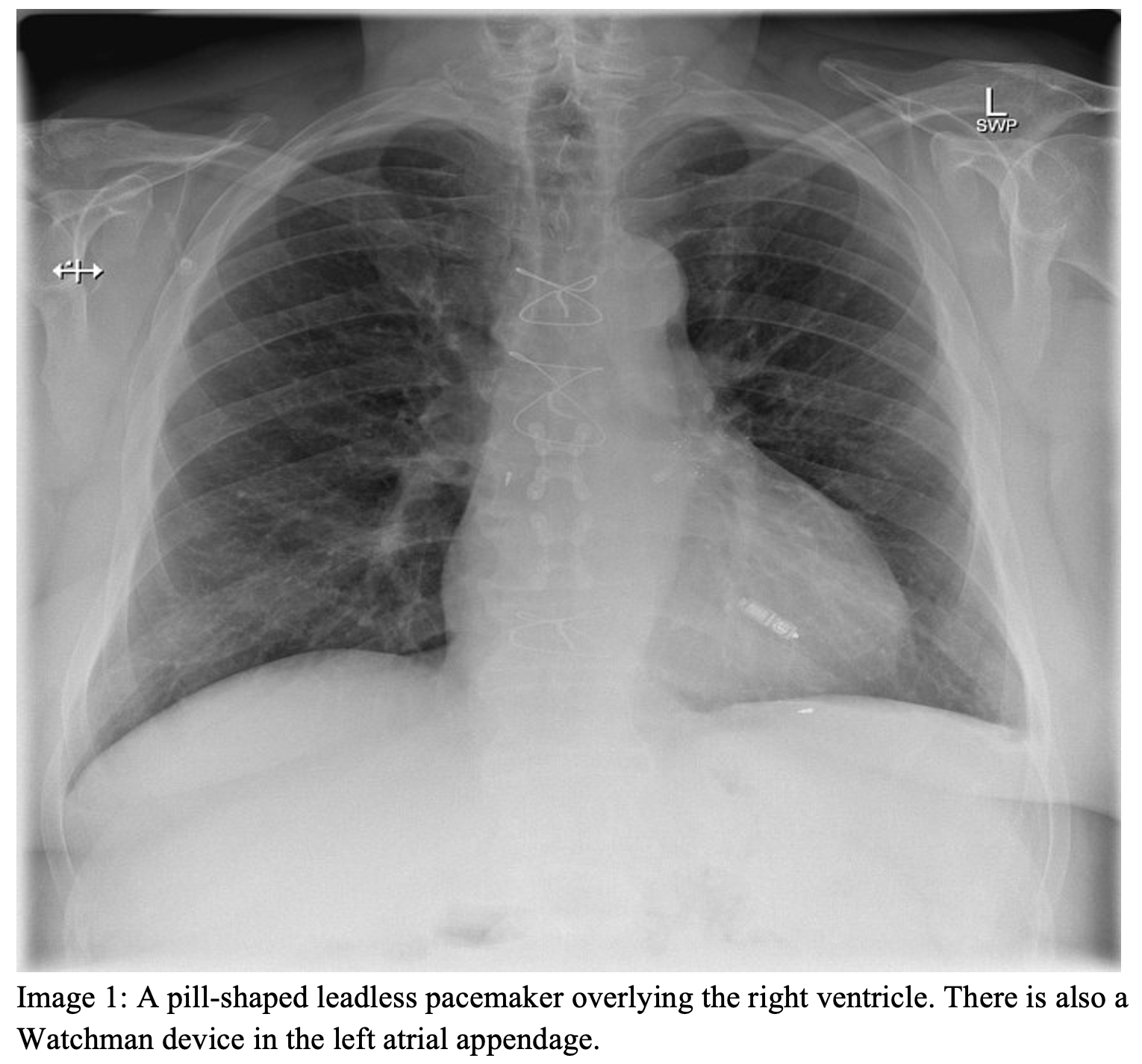
An 81-year-old male presents with a heart rate of 31 and a blood pressure of 81/47. He reports feeling lightheaded and chest pain. He recently had a pacemaker placed for having a “slow heart rate” but is unsure of information beyond that. On exam, there is no pacemaker noted on the chest. X-ray demonstrates a small radiopaque object overlying the heart.
Question: What kind of pacemaker does the patient have?
Answer: Leadless Cardiac Pacemaker
Background:
- Leadless cardiac pacemakers (LCPs) are increasingly used in the place of traditional pacemakers (TPMs) in the treatment of brady-arrhythmias.[1]
- They are associated with reduced rates of inpatient mortality, a better quality of life with less limitations related to discomfort.[2]
- Most LCPs are MRI-compatible, but confirm with the manufacturer.[3]
- The first LCPs perform single-chamber ventricular pacing, being implanted in the RV and largely used for atrial fibrillation with low ventricular response and cases where a high amount of pacing was not expected such as sinus node disease, syncope, or frequent paroxysmal AV blocks.[4]
- Newer LCPs may perform atrioventricular-synchronized pacing, allowing for their use in patients with sinus and AV node disease.[5]
Comparison with Transvenous Pacemakers:
- TPMs have a subcutaneous pulse generator placed in the chest wall and pacing leads that attach to the myocardium.
- LCPs are 90% smaller and require no subcutaneous generator pocket, eliminating the risk of pocket infections, lead fracture, hematoma, and lead dislodgment.[6]
- There is a lower risk of pneumothorax and dislodgement and decreased overall mortality with LCP compared to TPM.[7]
- The median projected longevity of LCP is 10-16 years, depending on the model and patients’ pacing dependency.[8]
- LCPs can be retrieved at the end of their life, though some may become encapsulated and left in place and another is implanted alongside the original.[9]
Evaluating the Pacemaker:
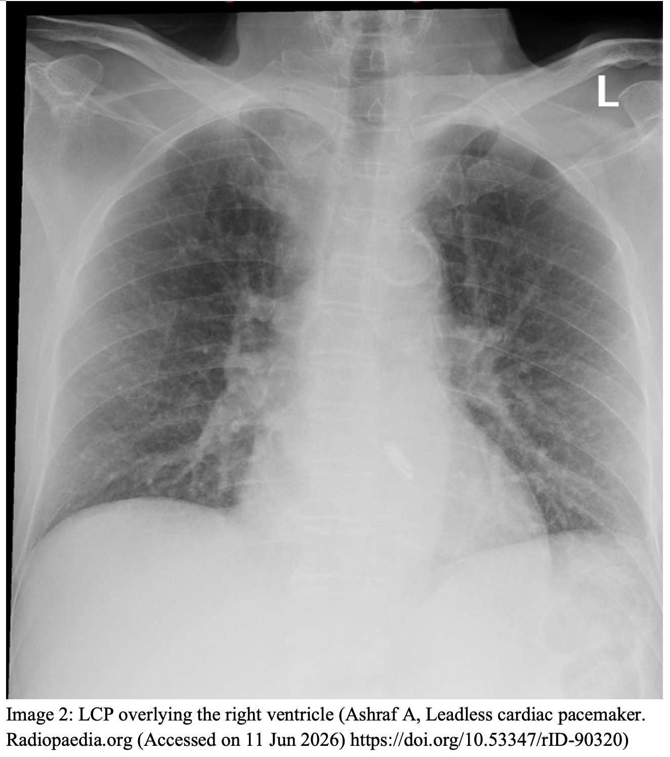
- On x-ray and CT, LCPs appear as linear radiopaque objects, usually in the RV wall.[10]
- Interrogation equipment is rarely readily available in all EDs, reach out to the device representative for guidance. The currently approved makers are Medtronic (Micra) and Abbott (Nanostim, Aveir).
- Nanostim was recalled in 2016 due to battery failures and is no longer used.
- Patients may be able to receive telemetry data via an application on their smart phone or have at-home equipment for transmitting data.[11]
- Leadless cardiac resynchronization therapy (CRT) is possible with leadless pacemakers but is not yet widely used.
- Ultrasound-based wireless communication from a submuscular transmitter to endocardial electrode for LV pacing, and with existing RV pacing.[12]
Types of LCPs:
- Right Atrial Leadless Pacemaker (Abbott Aveir AR).[13]
- Indication: sinus node dysfunction with preserved AV nodal conduction
- Location: right atrial appendage along the anterior-lateral wall.
- Able to sense intrinsic p waves, and if detected, will inhibit itself. If no p wave is detected, it will deliver a pacing impulse followed by a p wave
- EKG: p wave morphology may look slightly different than normal
- II, III, aVF – p waves are upright and positive, longer in duration and diminished in amplitude when compared to intrinsic p waves.
- V1 – p waves negative or biphasic (initially positive)
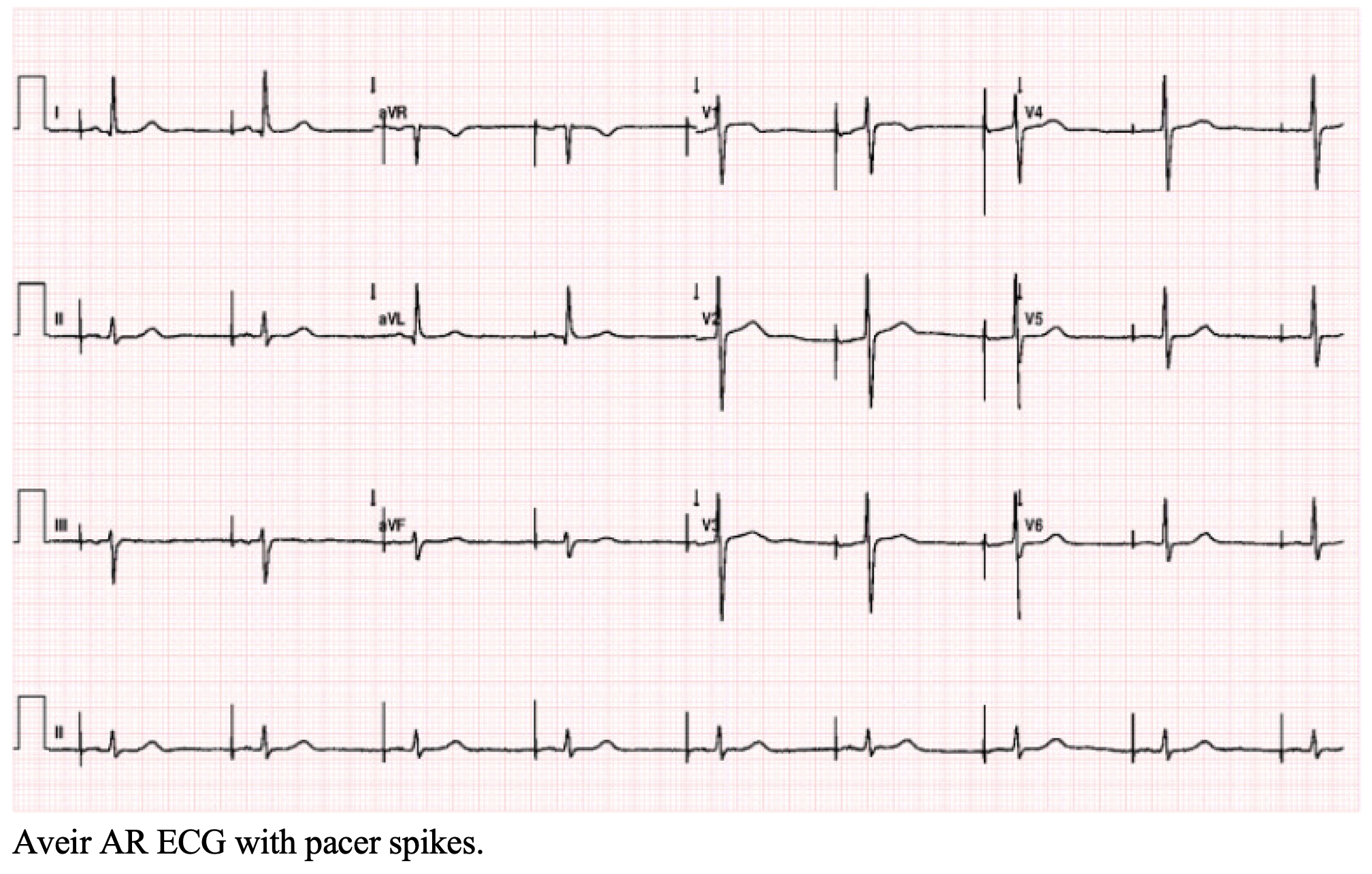
- Right Ventricular Leadless Pacemaker – single chamber system (Micra VR/VR2, Aveir VR) [14]
- Indication: Bradycardia needing only ventricular pacing
- Location: Most commonly in the mid-septum (there are fewer cases of pacemaker induced cardiomyopathy and ventricular perforation when here)
- EKG: Typically left bundle branch block (LBBB) morphology.
- QRS duration depends on placement location.
- Can function in VVI where it paces ventricular beats, senses ventricular beats, and inhibits itself in response to sensing; can also be set to be VVIR mode which allows rate-responsiveness during physical activity
- Right Ventricular Leadless Pacemaker – single chamber system with atrioventricular (AV) tracking (Micra AV and Micra AV2)
- Indication: Frailty, kidney disease (especially those on dialysis), <10 years life span, difficult access for transvenous pacemakers. [16]
- Location: Mid-high intraventricular septum
- EKG: Intrinsic p waves from the patient followed by a wide QRS.
- The pacing spike will likely be absent given the low energy needed to stimulate the beat
- Uses internal accelerometers to mechanically track atrioventricular conduction (as opposed to electrical tracking in traditional pacemakers). [17]
- AV synchrony is most consistent with sinus rates under 80 beats per minute (bpm).
- They have an upper tracking rate. At faster sinus rates, the ability to track atrial activity degrades so there may be loss of p-QRS coupling and reverts to VVI or VVIR based on settings.
- AV synchrony is most consistent with sinus rates under 80 beats per minute (bpm).
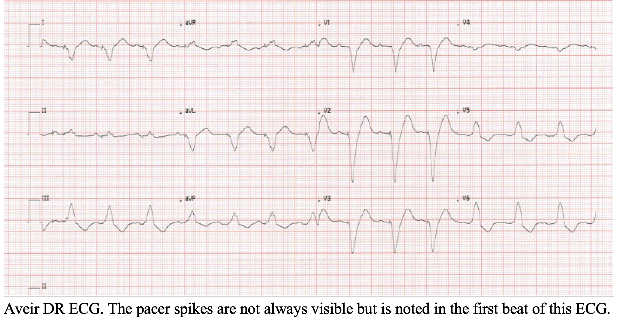
- Dual-chamber leadless pacemaker (Aveir DR) [18]
- Indication: Significant sinus node disease with an anticipated high pacing burden, or significant AV nodal disease.
- Achieves 98% synchrony including >95% synchrony across all heart rate ranges. [18]
- Location: One device in the right atrium, one in the right ventricle
- ECG: there may or may not be visible pacemaker spikes.
- The standard mode is DDD where both the atria and ventricles are paced, both are sensed, and they can be either inhibited or triggered.
- An atrial leadless pacemaker and a ventricular leadless pacemaker communicate via extremely low electrical impulses transmitted through blood and myocardial tissue (Knops et al, 2023).
- Indication: Significant sinus node disease with an anticipated high pacing burden, or significant AV nodal disease.
Complications:
- Physical complications related to the LCP [10,19–21]
- Dislodgement and cardiac perforation can be evaluated on x-ray as a change in location of the device, but are much better evaluated on echocardiogram and CT.
- Perforation symptoms can include chest pain, shortness of breath, syncope, up to tamponade. Failure of the pacemaker is common with dislodgement and perforation.
- Emergent consultation with the interventionalist (cardiology or surgery) if concerned.
- Vascular complications such as hematoma, pseudoaneurysm, arteriovenous fistula, acute limb ischemia, and pulmonary embolism should be evaluated with CT angiography imaging.
- Deep vein thrombosis may occur at the access site related to LCP placement; risk is increased in hypercoagulable patients or those with prolonged procedures.
- This should be diagnosed with ultrasound imaging.
- Dislodgement and cardiac perforation can be evaluated on x-ray as a change in location of the device, but are much better evaluated on echocardiogram and CT.
- Failure of the LCP
- Battery failure can occur as with standard pacemakers, and is a common reason for implantation of an alternate pacer. [13,14]
- Similar to traditional pacemakers, leadless pacemakers can suffer from a failure to pace or a failure to sense. This can lead to ineffective or inappropriate pacing and has to do with largely the contact point and fixation with the myocardial tissue.[22,23]
Managing the Malfunctioning Leadless Pacemaker:
- If the LCP fails, transcutaneous pacing can temporize the patient and a transvenous pacemaker may be placed when indicated.[25,26]
- Cardioversion may induce a power-on reset mode and interrupt device function, but the device should resume normal function quickly and retain programmed settings.[27]
- End of life pacemaker management is still not well defined. Options are:
- Removal of the non-functioning pacemaker and implanting a new one or
- Placing an additional leadless pacemaker adjacent to the existing one.[28]
- Consultation with cardiology and device representatives can assist in evaluating the LCP.
- Magnets:
- Medtronic LCPs are not affected by pacemaker magnets; they must be reprogrammed if malfunctioning.
- Abbott LCPs (as long as magnet mode is not set to off) changes to an asynchronous node and paces at 100ppm for eight cycles, then either 90ppm or 65ppm depending on the pacemaker. When the magnet is removed, the LCP reverts to the previous program within 5 seconds.[29,30]
Pearls:
- Leadless cardiac pacemakers may be more difficult to see on chest x-ray compared to traditional pacemakers.
- There are various types of LCPs with different capabilities. The specifics for each patient should be available from their pacemaker card, previous charts, or from the manufacturer.
- If the LCP fails, transcutaneous or transvenous pacing can be done with it in place.
- LCPs are often left in place at the end of their life; new ones can be implanted alongside the old one.
- The efficacy of pacemaker magnets depends on the specific device and manufacturer.
 A 72-year-old man undergoes implantation of a leadless ventricular pacemaker for symptomatic bradycardia. What ECG pattern is he most likely to have after implantation?
A) Atrial paced rhythm with normal PR interval
B) Narrow QRS complexes with absent pacing spikes
C) Right bundle branch block morphology with ventricular pacing spikes
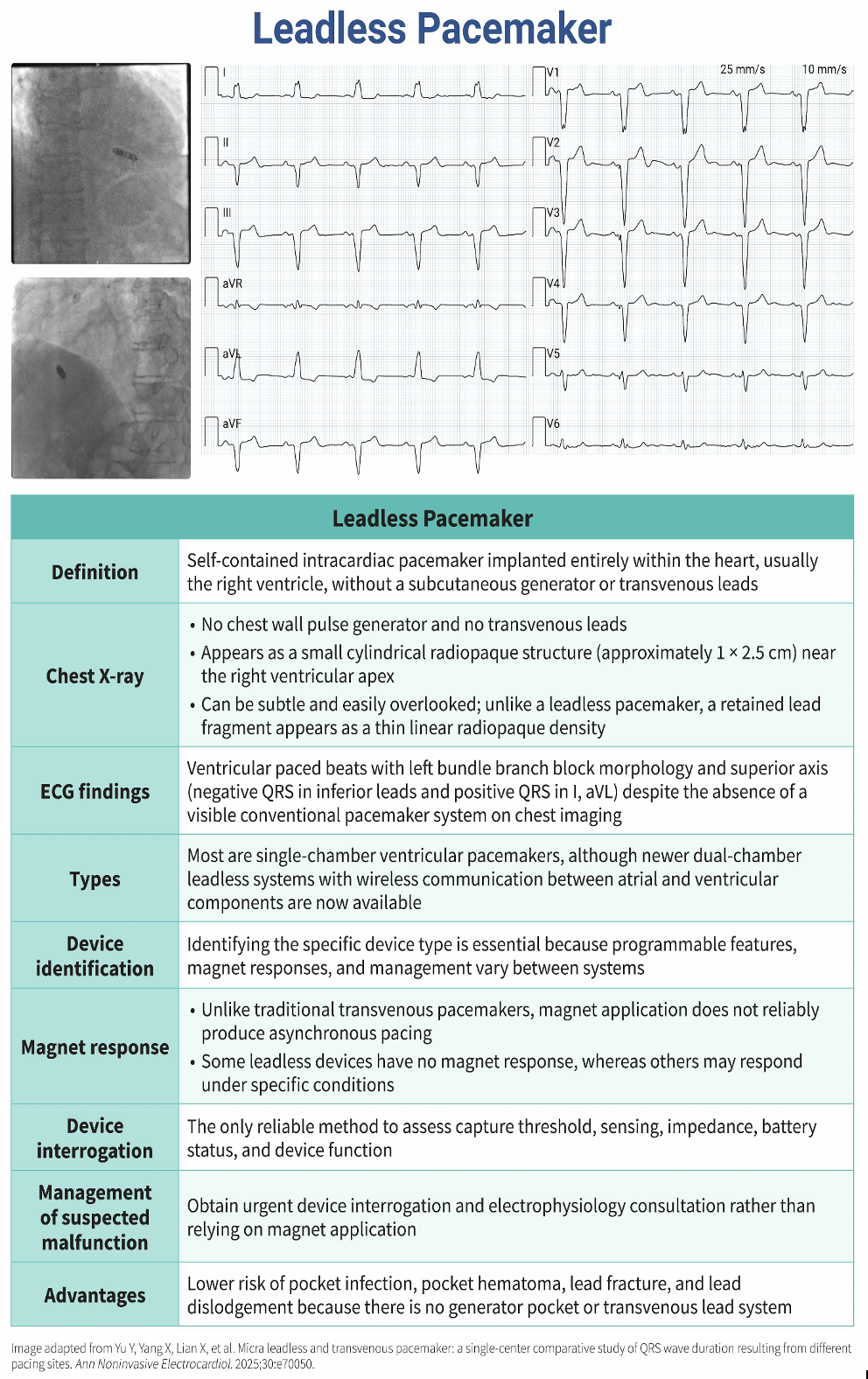
D) Ventricular paced rhythm with left bundle branch block pattern and superior axis
Answer: D
Leadless ventricular pacemakers are implanted directly within the right ventricle, most commonly at the right ventricular apex or midseptum. When ventricular pacing occurs from the right ventricle, electrical activation spreads through the myocardium rather than the native His-Purkinje conduction system, resulting in a characteristic paced ECG pattern.
The ventricular-paced QRS complex demonstrates left bundle branch block morphology because the right ventricle is activated first, followed by delayed activation of the left ventricle across the interventricular septum. This produces a broad QS or rS complex in lead V1 and broad monophasic R waves in the lateral leads.
When the device is positioned at the right ventricular apex, the paced depolarization wavefront usually travels superiorly, generating a leftward (superior) QRS axis with predominantly negative complexes in leads II, III, and aVF and positive deflections in leads I and aVL.
Pacing stimuli are generated by leadless pacemakers, and pacing spikes may be visible on surface ECG. However, because the device is entirely intracardiac, the spikes are often very small and may be difficult to identify or occasionally appear absent.
Atrial-paced rhythm (A) is not seen with leadless ventricular pacemakers. It paces the ventricle rather than the atrium (in single-chamber devices). Even the dual-chamber pacemaker uses a separate atrial component.
A narrow QRS without pacing spikes (B) is seen during atrial pacing. Ventricular pacing produces wide QRS complexes (typically > 140 msec), not narrow complexes.
Right bundle branch block morphology (C) during right ventricular pacing is uncommon and is more likely an artifact of lead placement or electrode positioning than the expected pattern. It would raise concern for inadvertent lead placement.
Further Reading:
- https://doi.org/10.1016/j.accpm.2025.101606
- https://pubmed.ncbi.nlm.nih.gov/41908186/
- https://pmc.ncbi.nlm.nih.gov/articles/PMC12127730/
References:
[1] Khan MZ, Nassar S, Nguyen A, et al. Contemporary trends of leadless pacemaker implantation in the United States. J Cardiovasc Electrophysiol 2024;35:1351–9. https://doi.org/10.1111/jce.16295.
[2] Yu M, Li YP, Shi DM, et al. Comparation of quality of life in Chinese patients undergoing leadless versus conventional pacemaker implantation. Clin Cardiol 2023;46:49–56. https://doi.org/10.1002/clc.23939.
[3] Soejima K, Edmonson J, Ellingson ML, et al. Safety evaluation of a leadless transcatheter pacemaker for magnetic resonance imaging use. Heart Rhythm 2016;13:2056–63. https://doi.org/10.1016/j.hrthm.2016.06.032.
[4] Malaczynska-Rajpold K, Elliot M, Wijesuriya N, et al. Leadless Cardiac Pacing: New Horizons. Cardiol Ther 2023;12:21–33. https://doi.org/10.1007/s40119-022-00288-0.
[5] Saleem-Talib S, Hoevenaars CPR, Molitor N, et al. Leadless pacing: a comprehensive review. Eur Heart J 2025;46:1979–90. https://doi.org/10.1093/eurheartj/ehaf119.
[6] Groner A, Grippe K. The leadless pacemaker: An innovative design to enhance pacemaking capabilities. JAAPA Off J Am Acad Physician Assist 2019;32:48–50. https://doi.org/10.1097/01.JAA.0000554750.85170.d4.
[7] Soleimani H, Dastjerdi P, Hosseini Mohammadi NS, et al. Safety and efficacy of leadless pacemakers: A systematic review and meta-analysis. Heart Rhythm O2 2026;7:598–618. https://doi.org/10.1016/j.hroo.2025.08.044.
[8] Leal MA, Sheldon T, Escalante K, et al. Device longevity of a leadless pacemaker family. Future Cardiol 2025;21:753–8. https://doi.org/10.1080/14796678.2025.2527466.
[9] Vouliotis AI, Roberts PR, Dilaveris P, et al. Leadless Pacemakers: Current Achievements and Future Perspectives. Eur Cardiol 2023;18:e49. https://doi.org/10.15420/ecr.2022.32.
[10] Conyers JM, Rajiah P, Ahn R, et al. Imaging features of leadless cardiovascular devices. Diagn Interv Radiol 2018;24:203–8. https://doi.org/10.5152/dir.2018.17462.
[11] Sundaram S, Chellappa N, Chaniary K, et al. Remote monitoring of leadless pacemakers using a novel conductive telemetry communication method. J Interv Card Electrophysiol Int J Arrhythm Pacing 2025. https://doi.org/10.1007/s10840-025-02095-8.
[12] Elliott MK, Sidhu BS, Mehta VS, et al. The importance of leadless pacemaker positioning in relation to subcutaneous implantable cardioverter-defibrillator sensing in completely leadless cardiac resynchronization and defibrillation systems. Hear Case Rep 2021;7:628–32. https://doi.org/10.1016/j.hrcr.2021.06.006.
[13] de Voogt WG, van Mechelen R, Scheffer M, et al. Electrocardiographic characteristics in low atrial septum pacing. J Electrocardiol 2005;38:166–70. https://doi.org/10.1016/j.jelectrocard.2004.10.005.
[14] Blank EA, El-Chami MF, Wenger NK. Leadless Pacemakers: State of the Art and Selection of the Ideal Candidate. Curr Cardiol Rev 2023;19:43–50. https://doi.org/10.2174/1573403X19666230331094647.
[15] Garweg C, Vandenberk B, Foulon S, et al. Leadless pacing with Micra TPS: A comparison between right ventricular outflow tract, mid-septal, and apical implant sites. J Cardiovasc Electrophysiol 2019;30:2002–11. https://doi.org/10.1111/jce.14083.
[16] Hrymniak B, Skoczyński P, Biel B, et al. Atrioventricular synchronous leadless pacing: Micra AV. Cardiol J 2024;31:147–55. https://doi.org/10.5603/CJ.a2023.0035.
[17] Steinwender C, Khelae SK, Garweg C, et al. Atrioventricular Synchronous Pacing Using a Leadless Ventricular Pacemaker: Results From the MARVEL 2 Study. JACC Clin Electrophysiol 2020;6:94–106. https://doi.org/10.1016/j.jacep.2019.10.017.
[18] Knops RE, Reddy VY, Ip JE, et al. A Dual-Chamber Leadless Pacemaker. N Engl J Med 2023;388:2360–70. https://doi.org/10.1056/NEJMoa2300080.
[19] Valente T, Bocchini G, Bigazzi MC, et al. First Multi-Detector Computed Tomography Evidence of Transcatheter Pacing System Migration and Embolization into the Pulmonary Vasculature. Korean J Thorac Cardiovasc Surg 2020;53:310–2. https://doi.org/10.5090/kjtcs.19.085.
[20] Hauser RG, Gornick CC, Abdelhadi RH, et al. Leadless pacemaker perforations: Clinical consequences and related device and user problems. J Cardiovasc Electrophysiol 2022;33:154–9. https://doi.org/10.1111/jce.15343.
[21] Hayashi T, Shishido KS, Moriyama NM, et al. Deep vein thrombosis after leadless pacemaker implantation. Eur Heart J 2022;43:ehac544.479. https://doi.org/10.1093/eurheartj/ehac544.479.
[22] Nomura T, Yamashita K, Nagashima M, et al. Predictive Role of Intraoperative Impedance in Midterm Pacing Threshold Elevation: Insights From Aveir VR Leadless Pacemaker Implantations. J Cardiovasc Electrophysiol 2025;36:1282–9. https://doi.org/10.1111/jce.16639.
[23] Tan JL, Epstein AE, Markman TM. Acute increase in pacing capture threshold and impedance post–leadless pacemaker implant with spontaneous resolution. Hear Case Rep 2024;10:453–5. https://doi.org/10.1016/j.hrcr.2024.03.016.
[24] Cossens M. Leadless cardiac pacemaker. Radiopaedia.org, Radiopaedia.org; 2018. https://doi.org/10.53347/rID-61188.
[25] Richter S, Döring M, Ebert M, et al. Battery Malfunction of a Leadless Cardiac Pacemaker: Worrisome Single-Center Experience. Circulation 2018;137:2408–10. https://doi.org/10.1161/CIRCULATIONAHA.117.033371.
[26] Nies M, Fluschnik N, Würger T, et al. Transient dysfunction of leadless pacemaker system after cardioversion. Hear Case Rep 2023;9:445–7. https://doi.org/10.1016/j.hrcr.2023.04.005.
[27] Tilz R, Nikorowitsch J, Traub A. Leadless Pacing and Cardioversion: Power On or Power Off? JACC Case Rep 2025;30:105485. https://doi.org/10.1016/j.jaccas.2025.105485.
[28] Beurskens NE, Tjong FV, Knops RE. End-of-life Management of Leadless Cardiac Pacemaker Therapy. Arrhythmia Electrophysiol Rev 2017;6:129–33. https://doi.org/10.15420/aer.2017:16:1.
[29] Özkartal T, Demarchi A, Caputo ML, et al. Perioperative Management of Patients with Cardiac Implantable Electronic Devices and Utility of Magnet Application. J Clin Med 2022;11:691. https://doi.org/10.3390/jcm11030691.
[30] Montandrau O, Untereiner O, Perin M, et al. Magnet Use for implantable cardiac devices: A Practical Guide. Anaesth Crit Care Pain Med 2025;44:101606. https://doi.org/10.1016/j.accpm.2025.101606.