
Author: Stephen Voyce, MD (Professor of Cardiology, Geisinger Community Medical Center) and Lloyd Tannenbaum, MD (EM Attending Physician, APD, Geisinger Wyoming Valley, PA) // Reviewer: Brit Long, MD (@long_brit)
Welcome back to ECG Pointers, a series designed to make you more confident in your ECG interpretations. This week, we feature a post from Dr. Tannenbaum’s ECG Teaching Cases, a free ECG resource. Please check it out. Without further ado, let’s look at some ECGs!
Dr. Sullivan, EM Chair: “Good morning, everyone, please take your seats, break is over! This week for the last lecture in Grand Rounds we have our STEMI conference. We’ll be joined by some of our interventional cardiology colleagues to run through two recent STEMI cases. Presenting the cases from the emergency room’s point of view will be Dr. Jackson and from interventional cardiology, we have Dr. Smith. These two have worked together for a very long time and they’re also married. They always put on a good, educational yet entertaining lecture. I know we’re a little short on time and this is the last lecture before lunch, so Dr. Jackson, the floor is all yours.”
Dr. J: Thank you, Dr. Sullivan. And thank you to Dr. Smith, our interventional colleague, for coming to conference today. Joint lectures like these are very beneficial for the residents to get a sense of the entire patient. We only get a quick snapshot of what happens while they’re in the ER. We really appreciate this view of what happens once they get rushed off by the cath team. Plus, it’s nice to hang out with you outside of work!
Dr. Smith: This doesn’t count as a date. We’re literally at the hospital, just a conference room, not a cath lab.
Dr. J: Uhhh. Right. Anyway, let’s take a look at the first ECG:
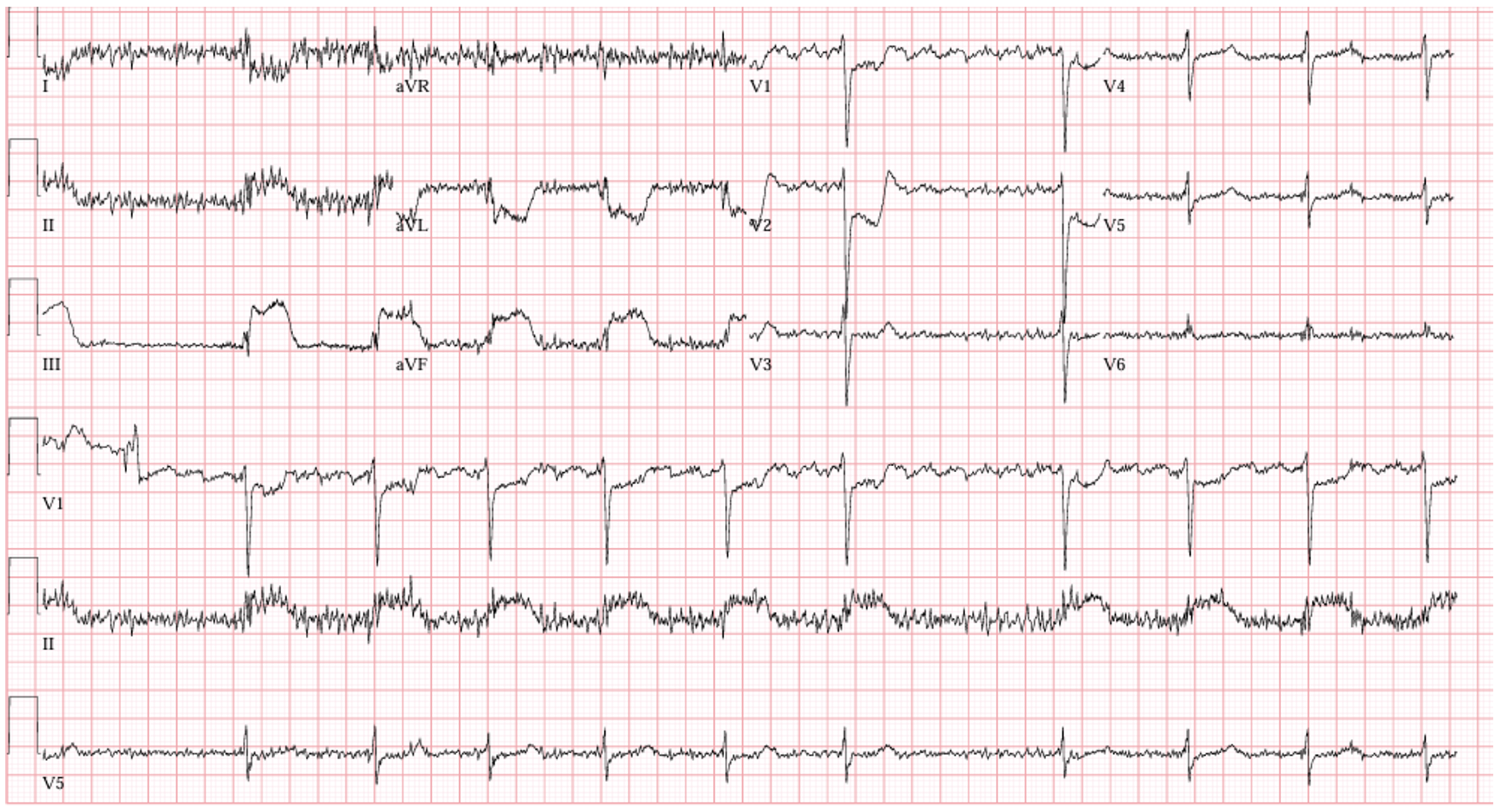
Dr. J: Residents, I’ll give you a few seconds to take a look and read this ECG before I go over what I see. This comes from patient JT who is an 85 year old male coming in with chest pain and shortness of breath. Long cardiac history including hypertension, coronary artery disease, high cholesterol, diabetes, afib, and probably more I’m forgetting right now. He described the chest pain as a crushing sensation, worse when he tried to walk around the house. He called EMS approximately 45 minutes after the pain started and arrived to the ER approximately 15 minutes later. Here’s my read of the ECG:
Rate: 60 beats per minute.
Rhythm: lots of artifact, a little tough to see. There are no obvious P waves that I can see plus the patient has a history of afib. With the constantly changing R-R interval, I’m ok going with afib here.
Axis: Probably normal. Up in I (I think, again artifact) and up in aVF.
Intervals: Narrow QRS complexes (<120 msec) and no PR interval.
Morphology: Here we go: Significant ST segment elevation in the inferior leads (especially III and aVF). Significant lateral depressions (I and aVL) and septal (V1/V2) depressions.
Final read: Massive Inferior MI
(** from a boards point of view, for a patient like this, we’d worry about the patient going into 3rd deg heart block, and look out for right ventricular involvement. This patient is likely preload dependent, and we should avoid nitro).
Dr. J: We activated the cath lab, and Dr. Smith, of course, agreed that this was a patient in need of emergent intervention. It’s so nice when you agree with me. Anyway, she took the patient upstairs and then…
Dr. Smith: Thank you, Dr. J. Before we get too far into the cath, I’d like to take a second to refresh what normal anatomy looks like on a cath and ignore your side comments. There are two main pictures I’d like you to keep in your head today. One that looks at the right coronary artery (RCA) and one that looks at left main coronary artery, left anterior descending (LAD), and left circumflex. Let’s do RCA first. This is what normal flow through the RCA looks like:

Dr. S: And this is flow through the left main mapped out. Notice that there’s still a wire in the LAD, so it’s a work in progress, but you can see the vessels pretty well:

Dr. S: Ok, so based on Dr. Jackson’s ECG and JT’s clinical story, we took him to the lab. Residents, based on the ECG, which vessel would you expect to be occluded?
Intern: Ma’am, it looks like an inferior STEMI, so I’d guess the Right Coronary Artery is blocked?
Dr. S: Excellent! Take a look at the cath images, this is before:
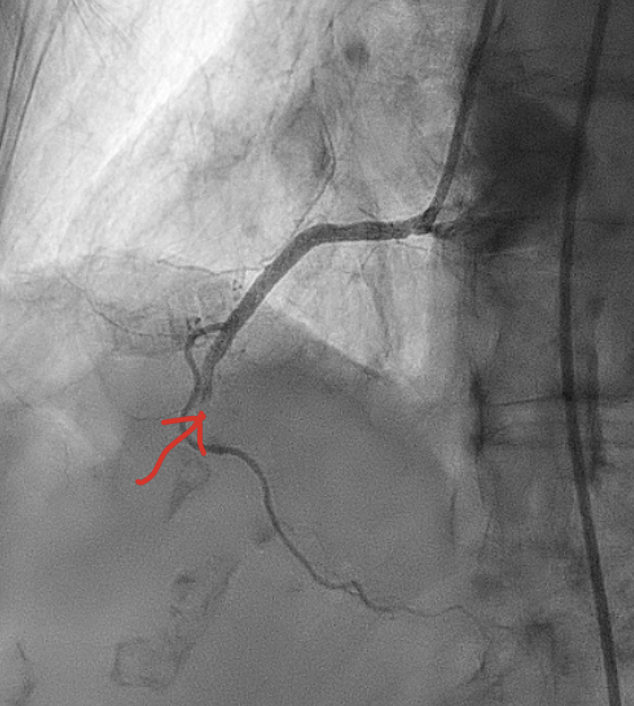
Dr. S: I added a red arrow to where the occlusion is. Notice how flow just abruptly stops. Now here’s an action shot from the cath with the balloon up:

Dr. S: And then finally, one with flow restored to the RCA:

Dr. S: To put it all together, patient JT had a Large dominant RCA with mid segment 100% occlusive heavily thrombotic stenosis status post successful PCI. Initially, he had TIMI 0 flow (remember, TIMI 0 means no flow) and, after stenting, had TIMI 3 flow (normal flow). He went on to make a full recovery and is now an active participant in our cardiac rehab program.
Dr. J: Thank you, Dr. Smith. Any questions on case 1? No? Ok then. Let’s move on to our second case. Again, take a look at this ECG while I tell you the story and then we’ll go through the findings:
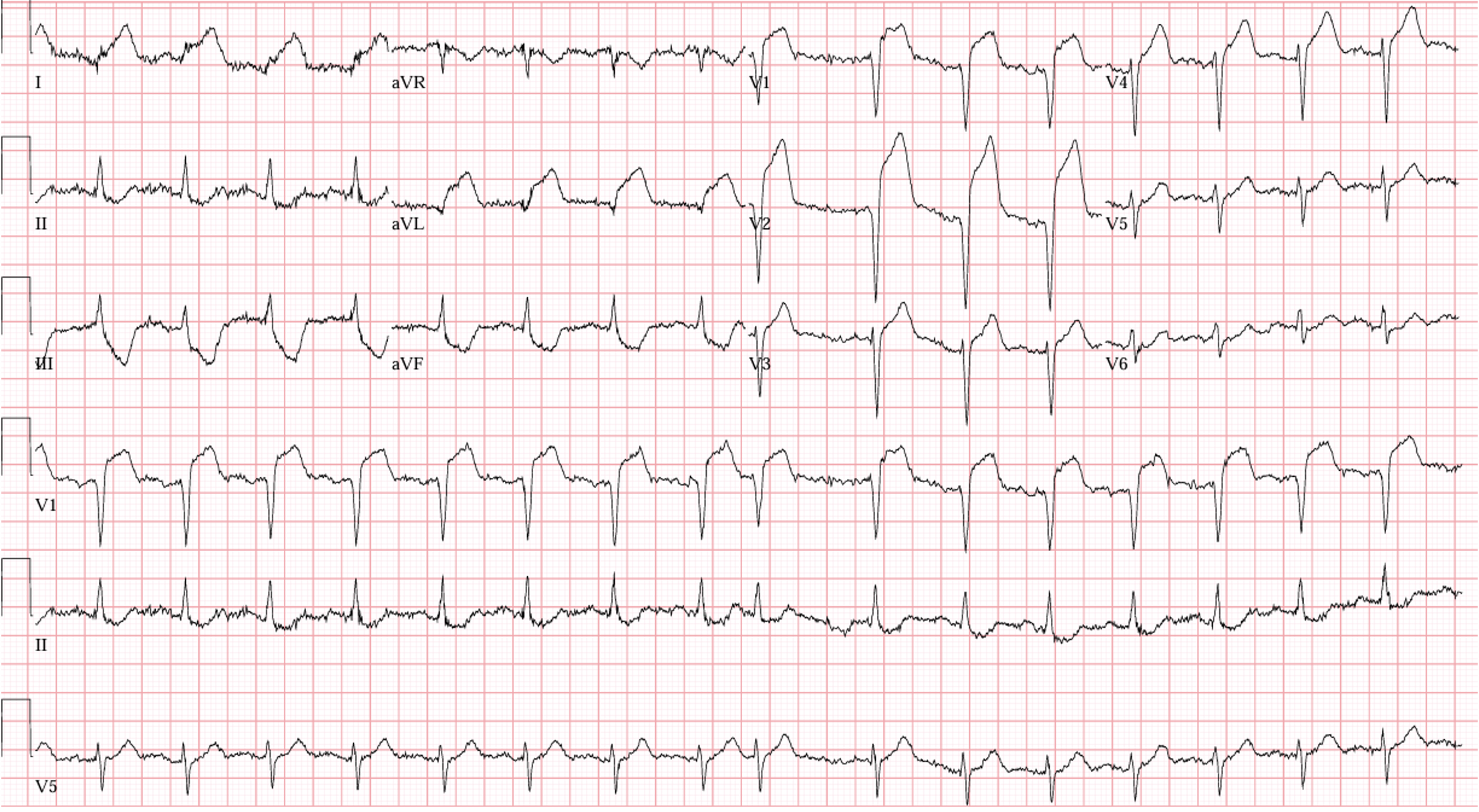
Dr. J: This patient, RF, is a 62 year old female who presented to the emergency room very unwell appearing. She was sweaty, vomiting, and looked like she was about to code. Here’s my ECG read:
Rate: around 100 bpm
Rhythm: Sinus right on the border of sinus tach.
Axis: Normal
Intervals: Narrow QRS, normal PR, Qtc looks ok.
Morphology: Significant elevation in the anterior/septal leads (V1, V2, V3, V4) with reciprocal changes inferiorly (best seen in III and aVF, also seen in II). There is also elevation in the lateral leads (I and aVL).
Final Read: Anteriolateral STEMI
Dr. J: Despite how ill she looked, her blood pressure was ok-ish. Sitting around 100/60. Right around now is when I called my wife about a new consult I had for her. She loves it when I call.
Dr. Smith: Yes, thank you so much for sending me yet another super stable patient to cath emergently. Am I allowed to block your number? Anyway, residents, based on this ECG, where do we expect the lesion?
Resident: That’s a tough question. I think based on the ECG, it’s probably an LAD lesion?
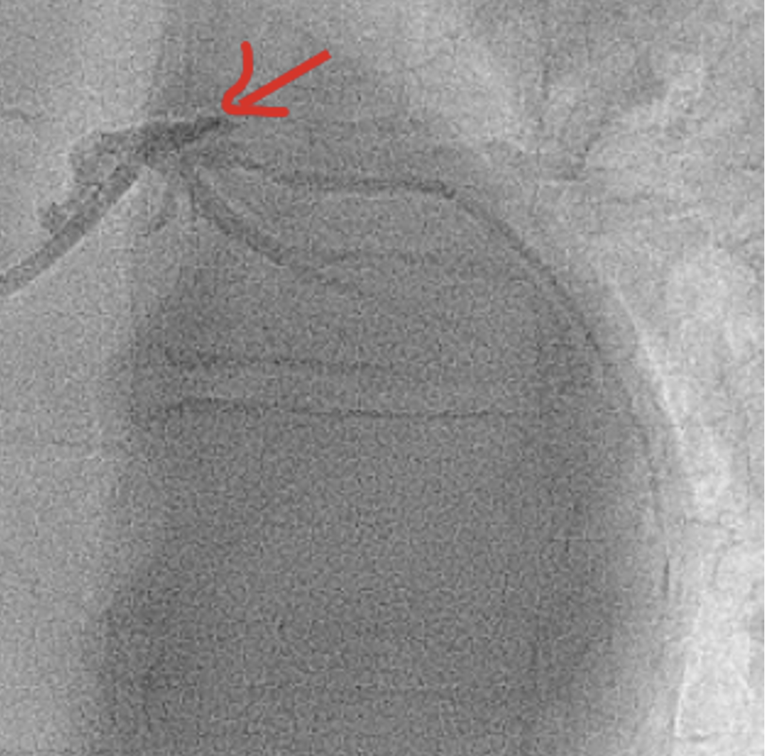
Dr. Smith: Correct, likely a proximal LAD lesion. This ECG is definitely a heart screaming for help. So, we took the patient upstairs and then it got a little exciting. This was the first shot that I got in the lab. I know, it’s a little tough to see, but I added a red arrow to highlight the pathology. That stump/abrupt stop is the LAD. It’s a little tough to see. Likely because the guy went into VF right as he got to the cath lab and it’s 100% occluded.
Resident: He went into VFib???
Dr. Smith: Yup, unfortunately. Luckily, we were able to shock him very quickly and he regained his pulse right away. Here’s a picture from after stenting, look at how much better that flow looks. A much happier heart:
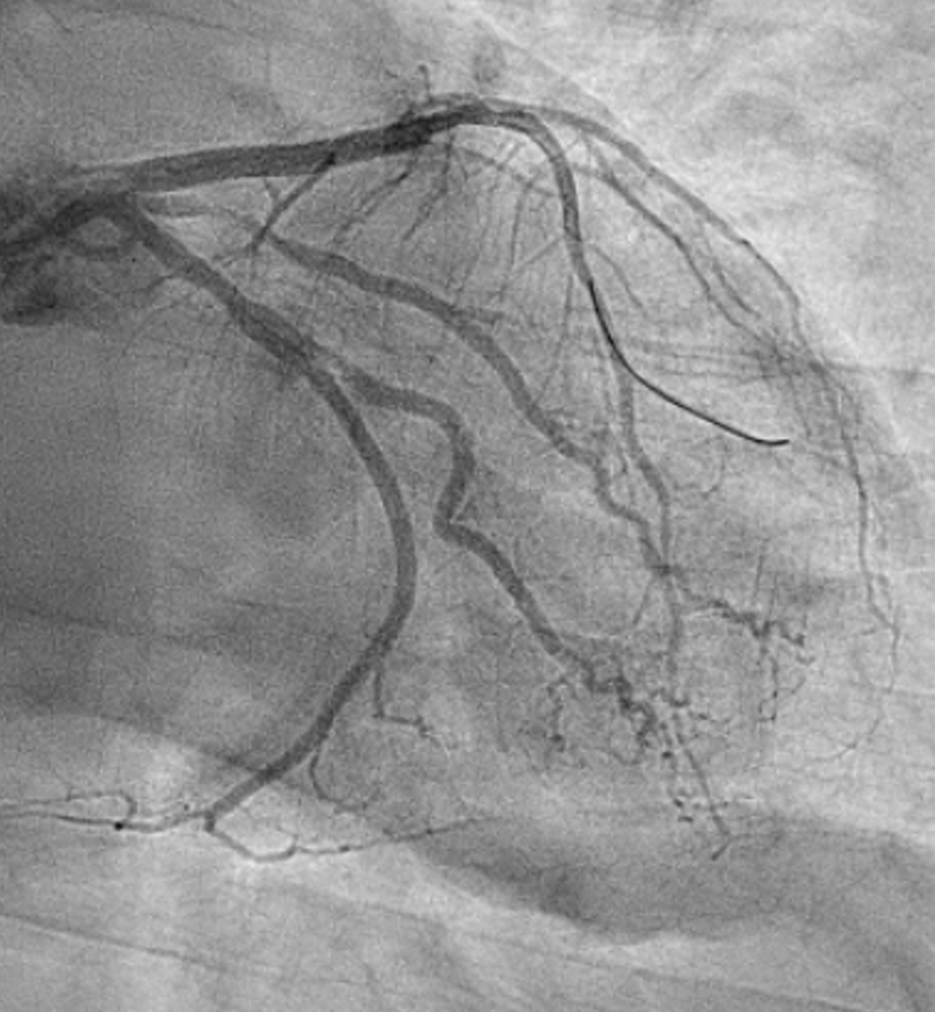
Dr. S: Ok, let’s wrap this case up. To summarize, RF had a Large caliber LAD with proximal 100% thrombotic occlusion. She had TIMI 0 flow initially and after stenting, TIMI 3 flow had been restored. Unfortunately, it seems like RF’s heart took a bit of a hit from the MI. She’s being followed closely in our heart failure clinic, her EF is less than 30 right now. We’ll see if it recovers, I’m hopeful that it will.
Dr. Sullivan: Thank you Drs. Jackson and Smith for sharing these cases with us. Dr. Smith, I don’t know how you put up with him. That concludes conference for today, see you all next week.
Case Wrap Up:
Since these cases are angiographically heavy, I ran them by one of my interventionalist friends and here were his thoughts:
Case 1:
- The cath film of the occluded RCA show a mid-vessel occlusion AFTER the 1st large RV marginal branch. This isn’t the typical angiogram of a hemodynamically significant RVMI, those usually show an occlusion proximal to the dominant RV marginal branches.
- The fact that this patient is in AF suggests they are probably on oral anticoagulation (OAC) which may complicate some decisions for intervention.
- Which site of arterial access? Radial is strongly preferred.
- Which OAC/antiplatelet regimen will be used during PCI?
- What is the antiplatelet/OAC regimen after PCI?
- The angiogram after revascularization shows the RCA to be large & dominant, giving off the posterior descending artery in the inferior IV groove & several posterior-lateral branches which is why you’re seeing the ST depressions in the precordial & lateral leads.
Case 2:
- The angiogram shows the initial occlusion very proximally, before the major septal perforator artery & that’s why you’re seeing septal STE (V1-2). There is also loss of R waves in the anterior leads suggesting that the occlusion has been present for a while & damage has already occurred.
- This is probably why the EF is reduced.