
Authors: Derek Fisher, MD (EM Resident, Geisinger Wyoming Valley, Wilkes Barre, PA); Lloyd Tannenbaum, MD (EM Attending, Geisinger Wyoming Valley, Wilkes Barre, PA) // Reviewed by: Brit Long, MD (@long_brit)
“Doc, I need you in room 7 NOW,” you hear Shannon, your charge nurse, tell you as you’re meeting a new patient across the ER. Something about her tone tells you it’s time to hustle over to room 7.
“Pardon me, I’ll be right back,” you tell the patient you were talking to as you walk out. “What’s going on Shannon?”
“EMS just brought in a guy to room 7 who looks terrible. Crushing chest pain, goes up to his jaw. It sure sounds like ACS to me. He’s profoundly sweaty and my sense is this guy is going to code soon if we don’t do something quick. Your intern is in there now talking to him. Here’s the EKG that EMS sent prehospital:
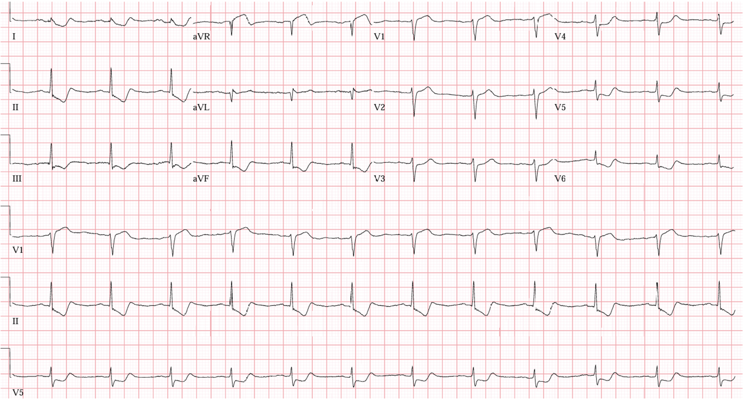
Rate: 70 ish beats per minute
Rhythm: sinus rhythm
Axis: normal
Intervals: Narrow QRS, QTc looks good, PR normal
Morphology: This looks very ischemic! ST segment elevation in aVR and V1, Diffuse ST segment depression in the inferior leads and anterior leads.
Final Read: This is an extremely concerning EKG for ischemia.
“Hello! What’s going on in here?” you say as you walk into the room.
“Oh good,” you hear the intern say. “I’m not sure what to do. This guy’s EKG looks bad, but I’m not sure if it meets STEMI criteria.
“Well, looking at him, I’m certainly concerned.” You say to the intern. “Looking at his EKG, I’m even more worried. Take a look at the elevation in aVR and those diffuse depressions. Tell me what you’re thinking about seeing those?”
“Well, we’ve seen ST segment elevation before, but it’s usually in the context of extreme tacychardia where there is diffuse strain on the heart. I don’t think this elevation is due to heart rate though. What else can cause this?” He asks.
“Great question! Let’s look a little bit at the history of aVR and talk about how it went from “the forgotten lead” to a major player for Left Main Coronary disease and then may have fizzled out a bit,” you tell the intern as he senses another ‘history of medicine’ lecture coming.
“But… This patient is in distress, shouldn’t we do something??” he asks.
“Young one, I’ve already activated the cath lab, talked to the interventionalists, updated the nurses, and placed all of the orders. The cath team is on their way down right now. That should give us plenty of time to talk. It all started in 2001…”
“Oh! At least this story starts in this millennia this time!” the intern quips.
“Silence! Now. As I was saying…”
Prior to 2001, there were some observational studies about elevations or depressions in lead aVR but it was mostly considered “The forgotten lead.”
In 2001, a landmark study in the Journal of the American College of Cardiology was published called, “Prediction of acute left main coronary artery obstruction by 12-lead electrocardiography. ST segment elevation in lead aVR with less ST segment elevation in lead V1.” The authors looked at the admission ECGs in 16 patients with acute left main coronary artery occlusion (LMCA), 46 with LAD occlusion, and 24 with RCA occlusion. What they found was that in the LMCA group, 14/16 (88%) had significant elevation in aVR, as comparted to 43% in the LAD group and 8% in the RCA group. Furthermore, if the elevation in lead aVR was greater than or equal to the elevation in lead V1, this was even more suggestive of an LMCA occlusion. This study was so impactful because it was one of the first studies to look at aVR using angiographic data to show its importance.
Then let’s fast forward a bit. There are a bunch of studies that came out between 2001 and 2013 that helped bolster support for aVR being a predictor of LMCA occlusion. In 2013, the next edition of the AHA/ACC STEMI guidelines were published and EKG nerds (like me) were excited to see elevations in aVR take its rightful place in the “STEMI Equivalent” category. But it didn’t happen. The 2013 guidelines mostly glossed over aVR and didn’t list it in the STEMI Equivalent category. Cardiology educators were surprised but some continued to teach elevations in aVR as a harbinger of LMCA occlusion, despite no formal guidelines to back them up.
And, I’m sure you noticed in the 2025 AHA/ACC ACS guidelines they also don’t really mention elevations in aVR, nor put elevations in aVR in the STEMI equivalent category. So, what happened? Do we still consider this indicative of LMCA occlusion? Or did it fizzle out? The answer is more complex than a simple yes or no. It’s more that the answer evolved.
Several studies came out that showed that STE in aVR is more indicative of global subendocardial ischemia, rather than isolated LMCA occlusion.
Notably, there is one in particular that we should talk about. In 2020, the Journal of Electrocardiology published a paper called “Is isolated ST segment elevation in Lead aVR associated with high grade coronary artery disease? A meta-anyalysis. The authors looked at 14 different studies to see if elevation in aVR was indicative of LMCA or 3 vessel disease and found that the sensitivity was only 40% and the specificity was 82%! And for just LMCA? Sensitivity was 39% and specificity was 86%. Not great numbers.
Ok, fine, let’s touch on one other meta-analysis published in 2020. This article looked at outcomes for patients with STE in aVR who were having ACS. Their question wasn’t “which vessel is the culprit; rather, what are the prognostic implications of elevations in aVR during ACS. After looking at over 7700 patients, they found that elevations in aVR during ACS is a very poor prognostic marker. These patients have higher rates of in-hospital mortality, reinfarction, heart failure, and 90-day mortality. Put simply, these patients are really sick!
Also, in February of 2021, JACC: Case Reports published a study called, “Electrocardiographic recognition of unprotected left main ST-segment myocardial infarction: looking beyond aVR.” [please note that an unprotected LMCA means that there is TIMI 0 flow, or no flow getting past the lesion]. In patients starting with a LMCA lesion with TIMI 0 flow (a very serious condition!) this paper published 2 cases and cited another paper where 7 patients had a total LMCA occlusion and no STE in aVR. As you can imagine, complete occlusion of LMCA is a BIG problem and many of these patients presented in cardiac arrest.
Ok, enough of me going on an on. What’s your take away from this? Is ST elevation in aVR always an LMCA occlusion? No! It’s likely indicative of diffuse subendocardial ischemia and these patients will likely be quite ill. They may need bypass grafting, therefore, they probably need an evaluation not just by interventional cardiology, but also by cardiac surgery. I’d keep a pretty close eye on them and get your consultants on board sooner than later.
Case Wrap Up:
Hey, Doc, while you were talking, the cardiology team already took the patient to the lab and sent me some pictures from the cath. Looks like it was an LMCA lesion this time:
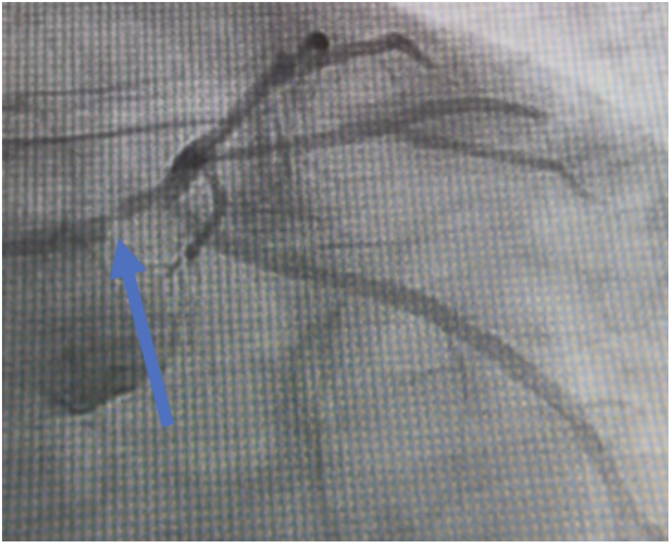
The blue line shows an LMCA lesion. Notice that, despite the lesion, there is distal flow, so this is NOT a TIMI 0 flow lesion.
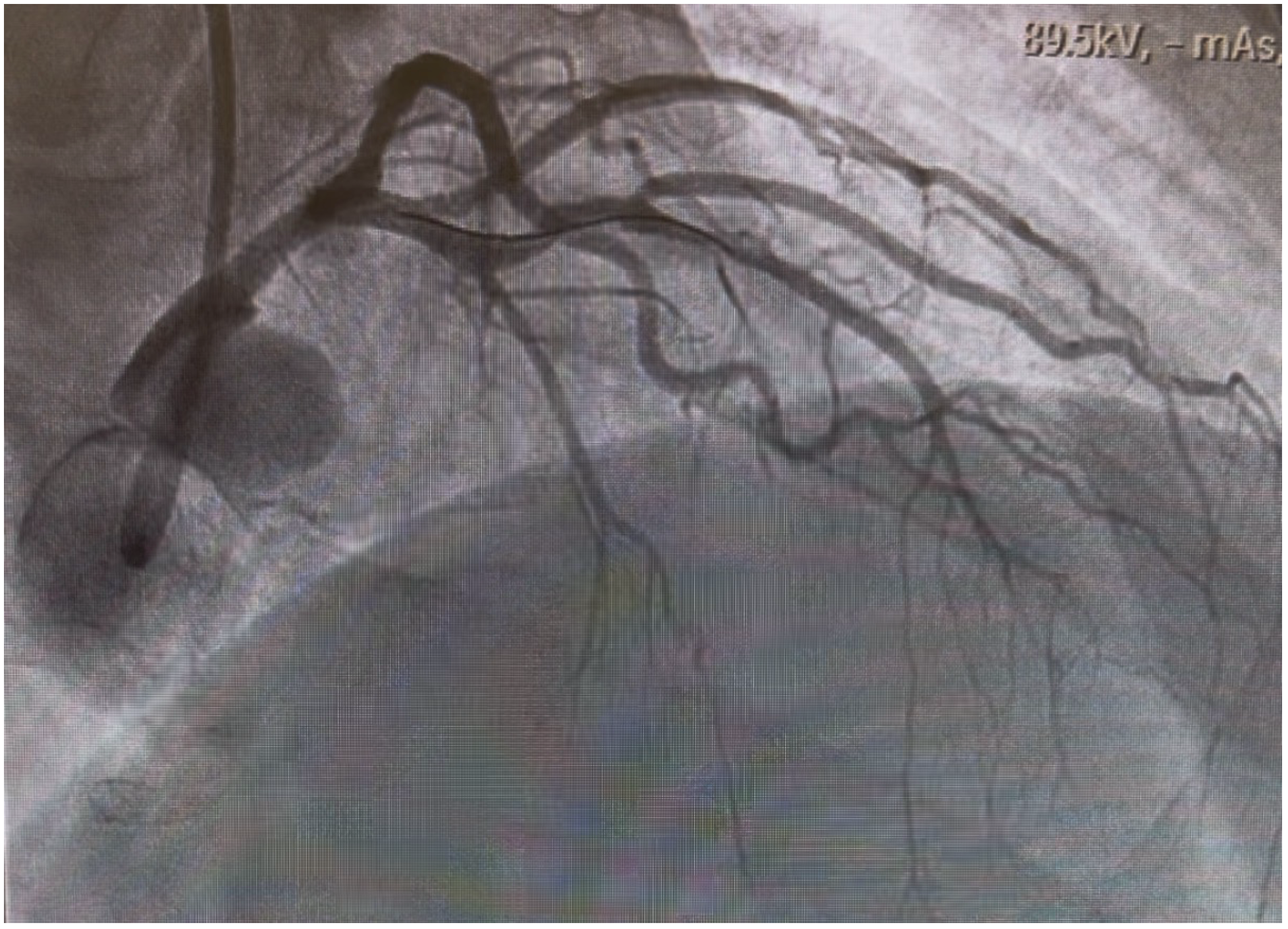
Here’s a shot after stenting, notice how much better those vessels look.
Recap:
- Lead aVR has gone from being forgotten to being one of the only harbingers of disease to shaking out somewhere in the middle
- For a while, elevations in aVR and, especially, elevations in aVR greater than elevations in V1 were taught to be an LMCA lesion
- Elevation in aVR should NOT be considered diagnostic of a LMCA lesion
- Nor is it considered a STEMI-Equivalent by the 2025 ACC/AHA ACS guidelines
- Additionally, the absence of elevation in aVR should not be mistaken to mean that there is no lesion in the LMCA
- Please note that patients with elevations in aVR will likely be quite sick and need a comprehensive cardiac work up, they may even need bypass grafting
- These patients have higher rates of in-hospital mortality, reinfarction, heart failure, and 90-day mortality.
- For some really interesting cases recapping elevations in aVR, take a look at: https://emergencymedicinecases.com/ecg-cases-st-elevation-avr-stemi-equivalent/