Episode 146: Pediatric Gastric and Midgut Volvulus
What is it?
- Twisting of the gastrointestinal (GI) tract on itself.
- May affect various segments; the stomach, small intestine, and colon.
How does it occur?
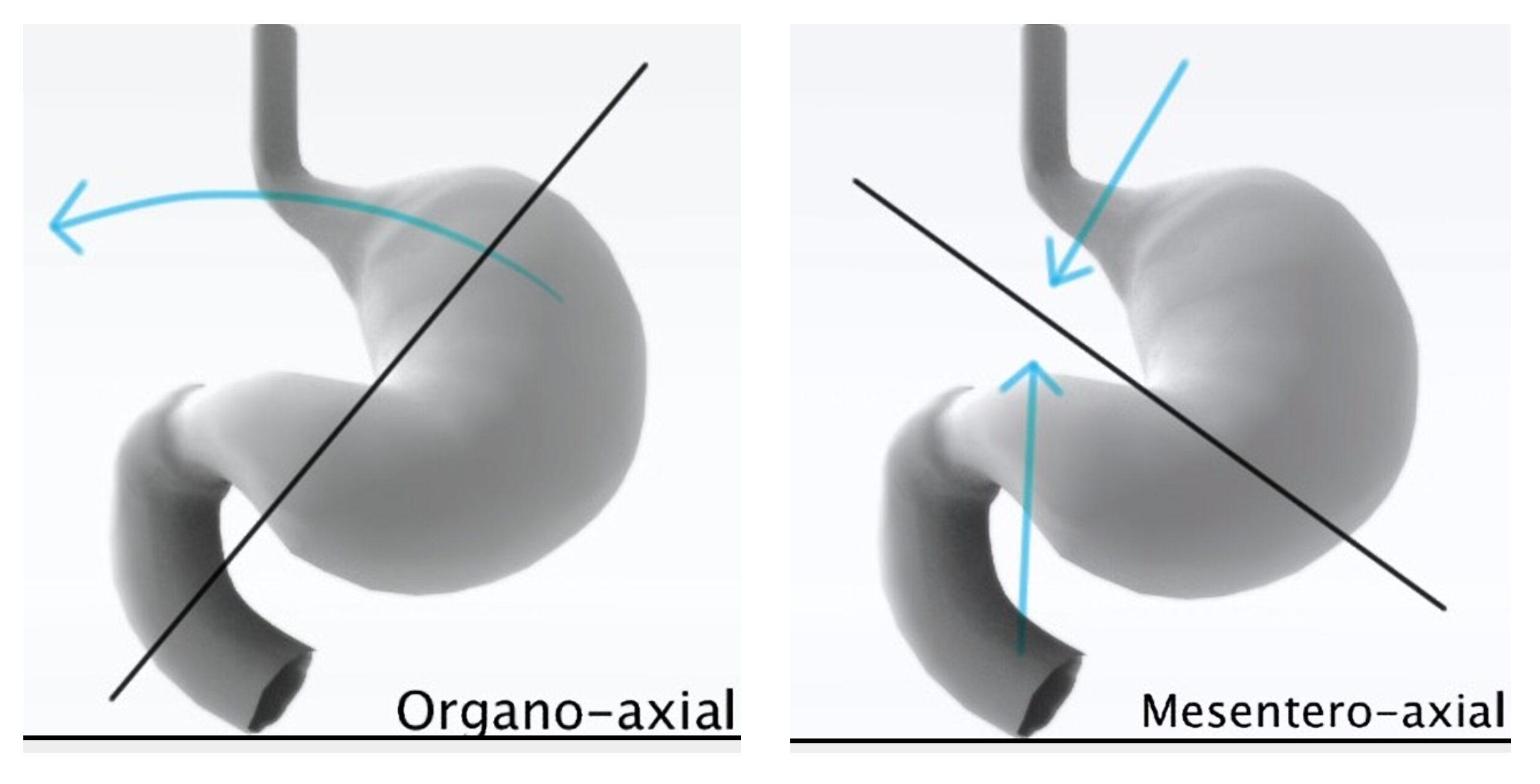
- Gastric volvulus is twisting of the stomach on itself by at least 180 degrees.
- Divided into several categories (axis versus cause).
- Axis: organoaxial form (most common, associated with the highest risk of strangulation and necrosis), mesenteroaxial, combined.
- Cause: idiopathic (2/3s) vs acquired/congenital abnormality. Both associated with abnormal stomach attachments and increased laxity in ligaments.
- Midgut volvulus is intestinal twisting.
- Most commonly associated with malrotation (disruption of embryologic sequence for the development, normal rotation, and fixation of the intestines which occurs from weeks 4-12 of gestation).
- Volvulus can lead to strangulation, necrosis, and perforation.
Figure 1. Types of gastric volvulus. Case courtesy of Maxime St-Amant, Radiopaedia.org, rID: 19257.
How common is this?
- Gastric volvulus is less common than midgut volvulus in the pediatric population.
- 581 cases of gastric volvulus reported between 1929 and 2007.
- Males and females affected equally; less than 20% of cases occur in children.
- Current mortality rates < 20%; the most common cause of death is strangulation resulting in necrosis with perforation. This occurs in up to 28% of patients with organoaxial gastric volvulus (15,16).
- Midgut volvulus: symptomatic neonatal malrotation occurs in 1 of every 6000 live births.
- Many of these patients with small intestine malrotation have some associated abnormality: congenital diaphragmatic hernia, congenital heart disease, gastroschisis, omphalocele, prune-belly syndrome, intestinal and esophageal atresias, biliary atresia, Meckel diverticulum, complex anorectal malformations, or Cornelia de Lange syndrome.
- Most cases of midgut volvulus occur in the neonatal/infant period (80% of cases in the first month following birth, 90% in the first year
- Mortality for midgut volvulus is up to 16%.
How do these patients present?
- Gastric volvulus:
- Classically presents with a triad – retching, epigastric distension, and difficulty passing a nasogastric tube. May be present in up to 70% of cases.
- Most common sign is retching or non-bilious vomiting.
- Patients often have progressive abdominal and epigastric distension and pain, but if the stomach is intrathoracic, distension and tenderness may not be present.
- Hematemesis and hiccups can occur.
- Midgut volvulus:
- Classic presentation is bilious emesis and initially a flat abdomen that then becomes distended.
- Over 90% of patients with present with vomiting. The emesis is usually bilious but it can be non-bilious.
- Abdominal pain is more common in older patients.
- Neonates and infants may have lethargy, fussiness, and decreased feeding.
- Hematemesis or hematochezia suggest ischemia.
- Severe ischemia -> evidence of sepsis with fever, tachycardia, hypotension) and peritonitis.
- Fluid loss from vomiting and third spacing from intestinal necrosis can lead to shock.
- Incomplete or intermittent volvulus may present with anorexia, nausea, recurrent abdominal pain, and growth failure; may occur over weeks to even months.
How do we make the diagnosis?
- Know what to look for!
- Bilious emesis in an infant is volvulus until proven otherwise. Consider volvulus if they have a condition that’s associated with malrotation and they’re vomiting, they have abdominal pain, or they’re unstable.
- If concerned, consult pediatric surgical specialist; may require transfer.
- If the patient is unstable or evidence of peritonitis (abdominal tenderness with rigidity or distension, hematemesis, hematochezia), resuscitate with IV fluids and broad-spectrum antibiotics, and consult pediatric surgical specialist.
- Imaging: X-ray, upper GI series, ultrasound (US).
- Gastric volvulus: start with X-ray of abdomen and chest, but upper GI series is definitive
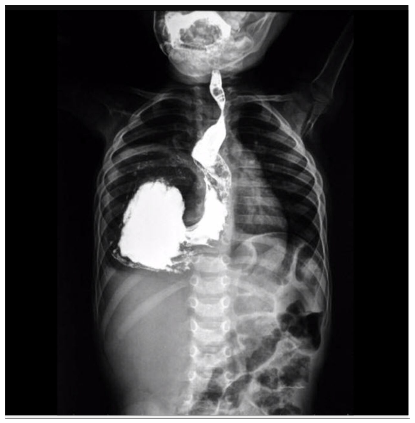
- X-ray organoaxial gastric volvulus: stomach horizontal with one fluid level.
- X-ray mesenteroaxial volvulus: two fluid volumes and a spherical stomach.
- Upper GI series is definitive diagnostic modality. In organoaxial volvulus, the stomach will be horizontal, and the lesser curvature is under the greater curvature with the pylorus oriented downward direction. In mesenteroaxial volvulus, upper GI series will show a pylorus above the GE junction.
- Gastric volvulus: start with X-ray of abdomen and chest, but upper GI series is definitive
Figure 2. Plain radiography with frontal view of the abdomen demonstrating the double bubble sign. https://commons.wikimedia.org/wiki/File:Radiograph_with_Double_Bubble_Sign.jpg.
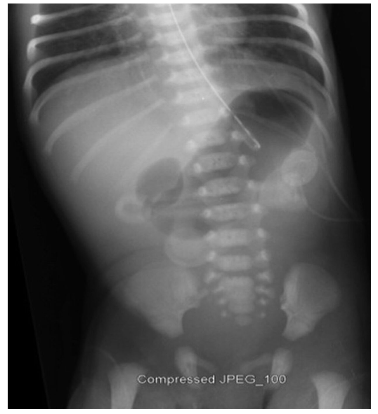
Figure 3. Organoaxial gastric volvulus, barium study. Case courtesy of The Radswiki, Radiopaedia.org, rID: 11449.
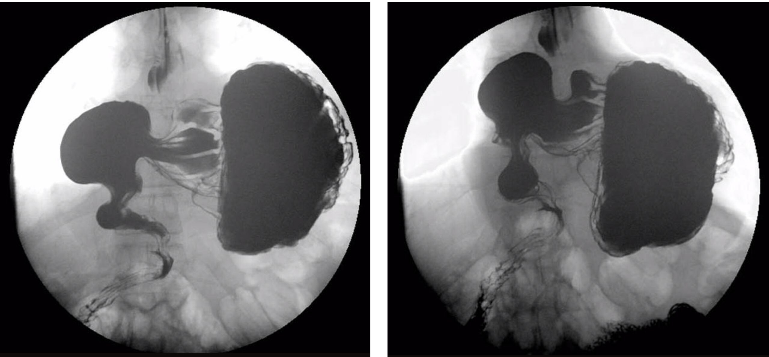
Figure 4. Gastric organoaxial volvulus. Case courtesy of Ahmed Abdrabou, Radiopaedia.org, rID: 24356.
-
- Midgut volvulus:
- Upper GI series with contrast is classically the gold standard for diagnosis.
- ACR appropriateness criteria recommendations are based on age.
- For patients within the first two days of birth, plain radiography is the recommended first-line modality: double-bubble or triple-bubble sign with no gas within the distal small intestine. A normal abdominal radiograph should not be used to exclude volvulus.
- For those older than two days, or if plain radiography is not diagnostic, upper GI series is the recommended modality. The sensitivity and specificity of upper GI series range between 85-95%.
- Findings on upper GI series: duodenojejunal junction displaced right and downward on the frontal view, duodenum with abnormal course on the lateral view, abnormally positioned jejunum on the right of the abdomen, dilated and fluid-filled duodenum, proximal small intestine obstruction, proximal jejunum spiraling down in the right or mid upper abdomen (corkscrew pattern), and mural edema and thick folds.
- Upper GI series demonstrates the highest sensitivity and specificity at pediatric centers with experienced radiologists.
- US is the other option and is often a first line modality.
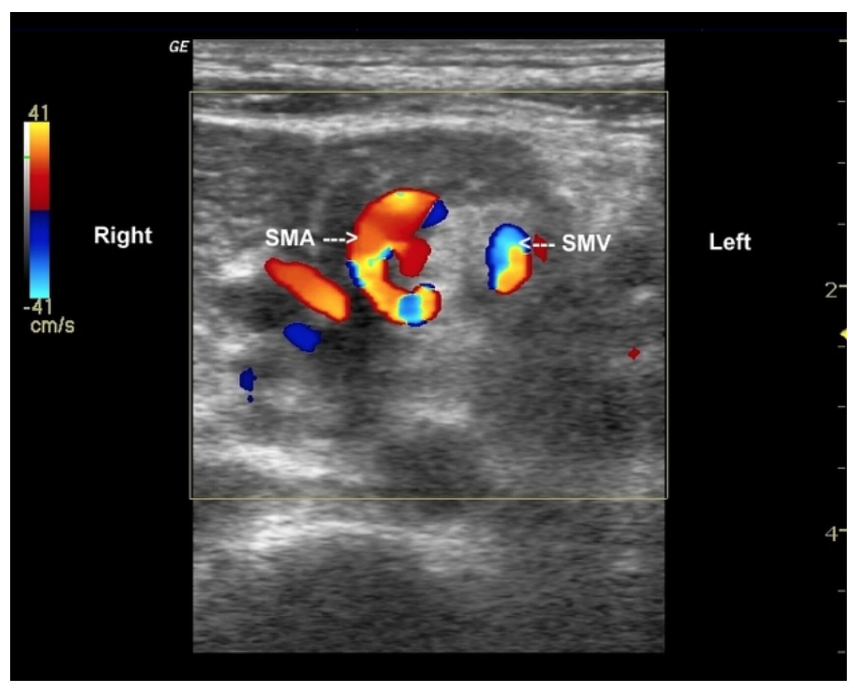
- May show reversal of the SMA and SMV and the whirlpool sign.
- Other findings: third portion of the duodenum anterior to the SMA, abnormal position of the cecum, hypoplastic uncinate process, a cutoff of the SMA, tapering/beaking of the third portion of the duodenum, dilated proximal duodenum, mesenteric edema, and ascites.
- Several studies have found a sensitivity of 83-100% for US diagnosis with a specificity ranging between 91-100%.
- The ACR states there is limited evidence for US diagnosis of volvulus, and they recommend against using a negative US to rule out midgut volvulus.
- If the US is nondiagnostic, then the patient needs the upper GI series.
- The choice between upper GI series and US should be based on local expertise and available resources.
- If concerned, consult surgeon and radiologist.
- May require transfer to the patient to a facility for imaging and pediatric surgeon.
- CT with IV contrast or laparoscopy are the final options.
- Midgut volvulus:
Figure 5. Corkscrew sign of malrotation with midgut volvulus in an infant. Spot film from a single contrast barium study demonstrates a corkscrew appearance of the fourth portion of the duodenum and the proximal jejunum and an abnormal position of the duodenojejunal junction. Case courtesy of Behrang Amini, Radiopaedia.org, rID: 35920.
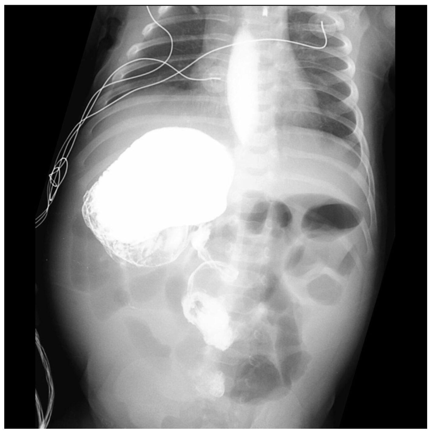
Figure 6. Contrast demonstrates a malplaced DJ flexure and a corkscrew appearance of the jejunum. This confirms both malrotation and volvulus. Case courtesy of Jeremy Jones, Radiopaedia.org, rID: 8078.
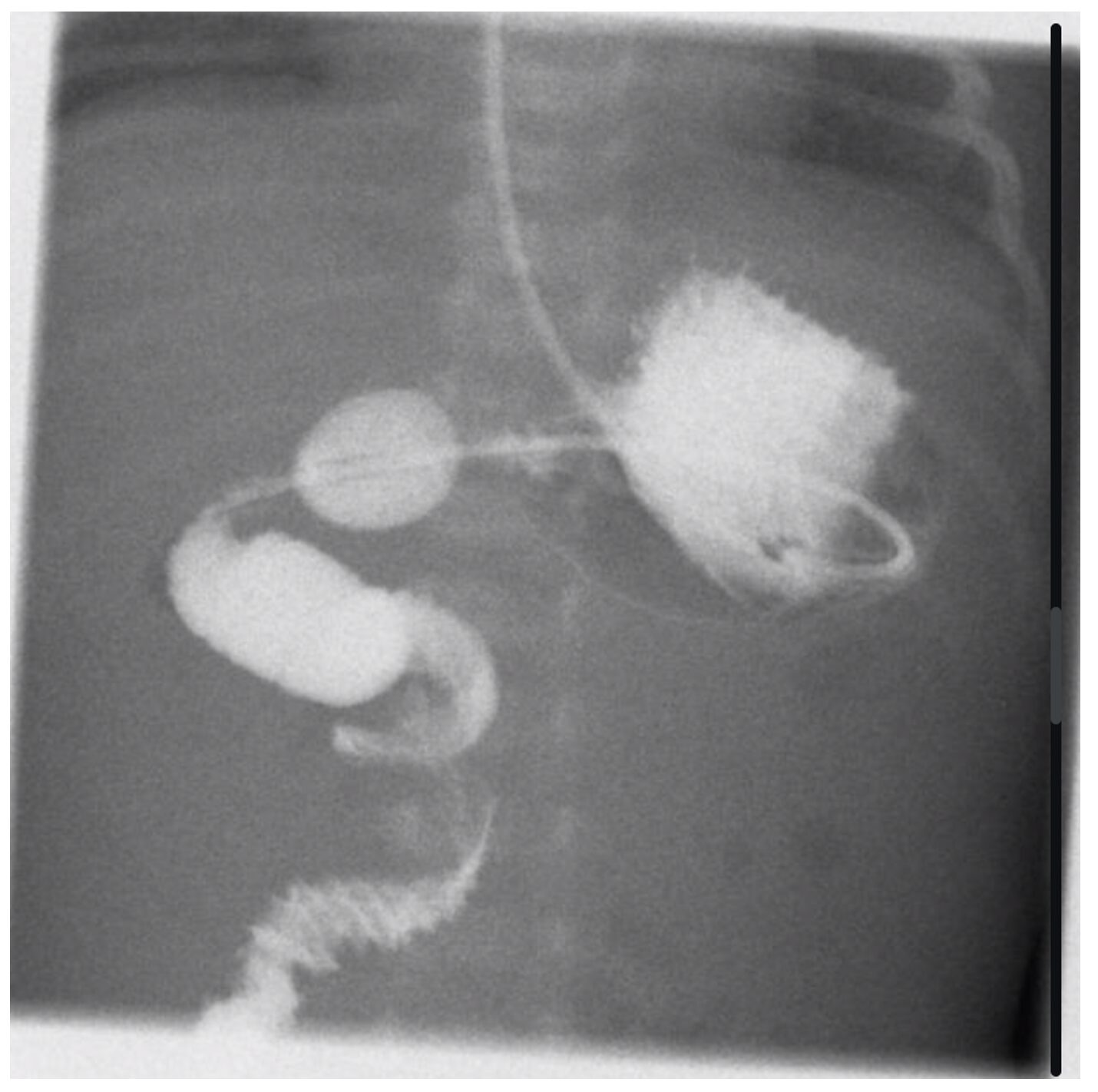
Figure 7. US with midgut volvulus. Grey scale and color Doppler US through the upper abdomen show whirling vessels and surrounding small bowel loops on transverse scan and corkscrew appearance on longitudinal scan. SMA-SMV relation is reversed. Case courtesy of KewalArunkumar Mistry, Radiopaedia.org, rID: 34790.
What’s the ED management of gastric and midgut volvulus in the ED
- All patients need emergent pediatric surgical consultation due to risk of ischemia and perforation.
- IV fluid resuscitation necessary due to volume loss and third spacing
- Antibiotics: piperacillin-tazobactam or ceftriaxone with metronidazole.
- Decompression of the GI system with an NG orogastric tube, but caution warranted due to risk of perforation, especially gastric volvulus.
- For gastric volvulus, surgical intervention includes reduction of the volvulus, assessment of gastric viability with resection of any gangrenous gastric portions, prevention of recurrence with gastropexy, and potentially fundoplication. There are minimally invasive approaches like laparoscopy and endoscopy.
- For midgut volvulus, surgical intervention is emergent laparotomy and detorsion of the volvulus, followed by the Ladd procedure. Involves widening of the mesentery base, placing the small intestine in a non-rotated position, and creating adhesions to keep the small intestine in place.
What are the long-term complications after surgically treated volvulus?
- Operative intervention successful in up to 90% of patients; mortality rate after surgery < 10%.
- Higher mortality rates in intestinal necrosis, prematurity, or associated anomalies, but rates are lower in otherwise healthy children and no intestinal ischemia.
- Risk of recurrence < 4% for patients undergoing laparoscopic intervention and < 2% for those undergoing the Ladd procedure
Summary:
- Volvulus is a dangerous condition associated with twisting of the stomach or small intestine on itself.
- Gastric volvulus most commonly presents with retching with or without nonbilious emesis, abdominal distension with pain, difficulty passing a nasogastric tube may be difficult.
- Midgut volvulus typically presents with bilious emesis in infants.
- Patients may experience hemodynamic compromise and peritonitis with perforation.
- If concerned, consult pediatric surgeon.
- Imaging includes radiography, US, and upper GI series, but testing needs to be based on local resources and experience.
- ED treatment includes resuscitation, broad-spectrum antibiotics, and emergent decompression.
References:
- Long B, Easter J, Koyfman A. High risk and low incidence diseases: Pediatric digestive volvulus. Am J Emerg Med. 2024 Aug;82:153-160.
- Langer JC. Intestinal Rotation Abnormalities and Midgut Volvulus. Surg Clin North Am. 2017 Feb;97(1):147-159.
- Garel C, Blouet M, Belloy F, Petit T, Pelage JP. Diagnosis of pediatric gastric, small-bowel and colonic volvulus. Pediatr Radiol. 2016 Jan;46(1):130-8.
- Do WS, Lillehei CW. Malrotation: Management of Disorders of Gut Rotation for the General Surgeon. Surg Clin North Am. 2022 Oct;102(5):837-845.
- Williams BS, Huynh TA, Mahmoud A. Gastric, Duodenal, and Small Bowel Emergencies. Surg Clin North Am. 2023 Dec;103(6):1097-1112.
- Adams SD, Stanton MP. Malrotation and intestinal atresias. Early Hum Dev. 2014 Dec;90(12):921-5.
- Shalaby MS, Kuti K, Walker G. Intestinal malrotation and volvulus in infants and children. BMJ. 2013 Nov 26;347:f6949.
- Filston HC, Kirks DR. Malrotation – the ubiquitous anomaly. J Pediatr Surg. 1981 Aug;16(4 Suppl 1):614-20.
- KIESEWETTER WB, SMITH JW. Malrotation of the midgut in infancy and childhood. AMA Arch Surg. 1958 Oct;77(4):483-91.
- Ford EG, Senac MO Jr, Srikanth MS, Weitzman JJ. Malrotation of the intestine in children. Ann Surg. 1992 Feb;215(2):172-8.
- Jain P, Sanghavi B, Sanghani H, et al. Congenital diaphragmatic hernia with gastric volvulus. Indian J Surg. 2007;69:260–263.
- Porcaro F,Mattioli G, Romano C. Pediatric gastric volvulus: diagnostic and clinical approach. Case Rep Gastroenterol 2013;7:63–68.
- Esposito F, Vitale V, Noviello D, et al.Ultrasonographic diagnosis of midgut volvulus with malrotation in children. J Pediatr Gastroenterol Nutr. 2014;59:786–788.
- Valsdottir E, Marks J. Volvulus: small bowel and colon. Clin Colon Rectal Surg. 2008;21:91–93.
- Milne LW, Hunter JJ, Anshus JS, Rosen P. Gastric volvulus: two cases and a review of the literature. J Emerg Med. 1994 May-Jun;12(3):299-306.
- Godbole P, Stringer MD. Bilious vomiting in the newborn: How often is it pathologic? J Pediatr Surg. 2002;37(6):909–11.
- Jackson R, Folaranmi SE, Goel N. Approach to the baby with bilious vomiting. Paediatrics Child Health. 2021;32(1):1–6.
- Raske ME, Dempsey ME, Dillman JR, et al. ACR Appropriateness Criteria Vomiting in Infants up to 3 Months of Age. J Am Coll Radiol. 2015 Sep;12(9):915-22.
- Expert Panel on Pediatric Imaging; Alazraki AL, Rigsby CK, Iyer RS, et al. ACR Appropriateness Criteria® Vomiting in Infants. J Am Coll Radiol. 2020 Nov;17(11S):S505-S515.
- Applegate KE, Anderson JM, Klatte EC. Intestinal malrotation in children: a problem-solving approach to the upper gastrointestinal series. Radiographics. 2006 Sep-Oct;26(5):1485-500.
- Ballesteros Gómiz E, Torremadé Ayats A, Durán Feliubadaló C, et al. Intestinal malrotation–volvulus: imaging findings. Radiologia. 2015 Jan-Feb;57(1):9-21.
- Tackett JJ, Muise ED, Cowles RA. Malrotation: Current strategies navigating the radiologic diagnosis of a surgical emergency. World J Radiol. 2014 Sep 28;6(9):730-6.
- Lampl B, Levin TL, Berdon WE, et al. Malrotation and midgut volvulus: a historical review and current controversies in diagnosis and management. Pediatr Radiol. 2009 Apr;39(4):359-66.