
Author: Seth Cohen, MD (@secohen11, EM Resident Physician, UTSW, Dallas, TX); Joshua Kern, MD (EM Attending Physician, UTSW, Dallas, TX) // Reviewed by: Alex Koyfman, MD (@EMHighAK) and Brit Long, MD (@long_brit)
Welcome to EM@3AM, an emDOCs series designed to foster your working knowledge by providing an expedited review of clinical basics. We’ll keep it short, while you keep that EM brain sharp.
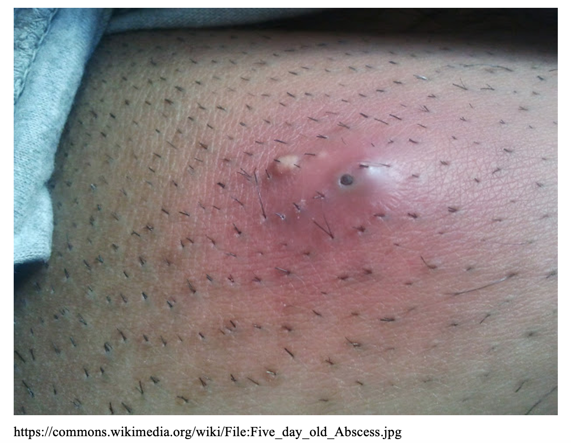
A 42-year-old male with no significant history presents to the ED with right buttock pain that started 3 days ago and has progressively worsened. He states that he has had this pain once before but can’t remember what was done. He reports feeling warm but denies any other symptoms. He noticed that the back of his underwear was wet this morning but wasn’t sure why.
Vital signs include HR 90 bpm, BP 148/100, RR 16, T 100.8F, and SpO2 96%. Examination is significant for a thin, disheveled male in no acute distress. His right buttock has a 5cm area of erythema with induration and fluctuance.
What is the most likely diagnosis?
Answer: Skin abscess
Background:
- Skin abscess may occur in healthy individuals with no predisposing conditions.
- Risk factors for abscess: Edema secondary to impaired lymphatic drainage or venous insufficiency, obesity, skin inflammation (eczema, radiation therapy, psoriasis), skin trauma (abrasion/laceration, ulcers, bites, intravenous drug use), immunosuppression, and pre-existing skin conditions1.
Pathophysiology:
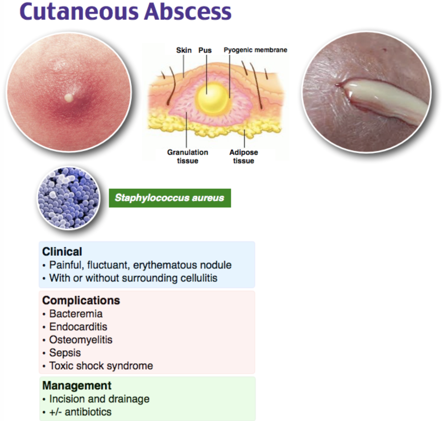
- An abscess is a collection of purulent material located within the dermis or subcutaneous space that is painful.
- Develops as a result of the body trying to fight off a bacterial infection. WBCs at collect at the site of infection, commonly at a hair follicle. The purulent material that develops is an accumulation of dead WBCs and bacteria.
- Most commonly caused by Staphylococcus aureus (either methicillin-resistant (MRSA) or susceptible (MSSA))2.
- Risk factors for MRSA-specific abscesses include recent hospitalization, living in a care facility, recent surgery, hemodialysis, HIV infection, intravenous drug use and sharing needles, recent antibiotic use, incarceration, military employment, sharing sports equipment. However, many MRSA carriers have no risk factors3.
- Skin abscesses can contain multiple bacterial species4.
Clinical Presentation:
- On examination, “fluctuant” and “indurated” are two words often used to describe abscesses. Fluctuant means a boggy-like feeling and indurated means firm and hardened.
- May or may not have surrounding cellulitis.
- Significant tenderness present
- Systemic symptoms including fever, chills, and lymphadenopathy are unusual4.
- If present, consider sepsis, necrotizing infection, and lymphadenitis.
Complications:
- If left untreated, complications include bacteremia and sepsis5.
- There are multiple types of abscesses aside from the commonly seen cutaneous abscess, with some having their own distinct complications and treatment protocols (only subcutaneous abscesses are discussed in this article):
- Breast abscess
- Different organ (liver, pulmonary, renal, splenic) abscesses
- Perirectal/perianal abscess
- Periodontal abscess
- Pilonidal abscess
- Bartholin’s cyst abscess
Evaluation:
- Abscess is a clinical diagnosis. If labs are obtained, they are nonspecific.
- CBC – possible leukocytosis
- ESR/CRP – possible elevation
- Ultrasound (picture below) can be used as a mass with heterogenous hypoechoic debris will be seen just beneath the skin. Assess for fluid collection and swirl within the collection.

- If patient has evidence of toxicity or systemic infection, obtain blood cultures, lactate, and renal function.
Differential Diagnoses:
- Cellulitis
- Erysipelas
- Folliculitis
- Hidradenitis suppurativa
- Lymphangitis
- Necrotizing fasciitis
- Epidermoid cyst
Management:
- Patients with fluctuant masses that are concerning for abscesses should undergo an incision and drainage and potentially receive antibiotic therapy.
- Standard incision and drainage steps:
- Prepare the surface of the abscess and surrounding skin with a cleaning agent such as iodine or chlorhexidine. Using sterile towels, drape around the abscess.
- Use lidocaine to perform a field block with a 25-30g needle by injecting the lidocaine in a ring around the abscess approximately 1 cm peripheral to the erythematous border. Do not inject local anesthetics directly into the abscess to avoid infiltration of the abscess down into the tissue or up toward you. Abscesses are acidic which causes lidocaine to lose effectiveness; IV hydromorphone hydrochloride or morphine sulfate may be utilized in addition. Wait approximately 5 minutes for the lidocaine to take effect.
- Before incising, make sure to use PPE to avoid self-contamination. Make a linear incision with a number 11 or 15 scalpel deep enough to penetrate the abscess. Cut the entire length of the abscess, as not making a wide enough incision could lead to inadequate drainage of the abscess.
- Once the abscess cavity has been reached, purulent material will begin to drain. Have 4×4 gauze on hand. Allow the purulent material to drain from the abscess. Gently probe the abscess with curved hemostats to break up loculations.
- Despite irrigation of the cavity commonly be referred to as standard practice, literature has shown that irrigation does not improve (or worsen) treatment success6.
- If packing material is warranted, (see considerations below), insert the packing in with hemostats or forceps. Dress the wound with sterile gauze and tape.
- The patient should be told to keep the wound clean, dry, and covered. Packing should be removed every one to two days and repacked until the cavity is resolved.
- Loop drainage technique: newly utilized incision and drainage that has been found to be equally as effective in adults, superior in children, and less painful.
- Make two small (4-5mm incisions) at the periphery of the abscess.
- Use a hemostat to break up the loculations, allowing the pus to drain.
- Irrigate the cavity with normal saline.
- Insert a vessel loop and pull through the other incision, tie ends together allowing for slack to avoid skin tension.
- Remove loop drain when the drainage has stopped, usually 7-10 days7.

- Antibiotic therapy is warranted if:
- Single abscess >2cm
- Multiple abscesses
- Surrounding cellulitis
- Immunosuppressed patient or patient with comorbidities
- Systemic symptoms
- Indwelling medical device is present
- High risk for transmission of staph aureus to others
- Antibiotic choices include (mg/kg is for pediatric dosing):
- TMP/SMX DS 1-2 tablets (5mg/kg) twice daily x7 days
- Clindamycin 450mg (10mg/kg) PO three times daily x7 days8
- In two different randomized trials, patients who received TMP-SMX resulted in higher cure rates after treatment than placebo (80.5-83 versus 69-73.6%)9, 10.
- No studies suggest that packing of a drained abscess (excluding abscesses greater than 5cm or in immunocompromised patients) reduces treatment time or recurrence of abscesses11, 12.
- Inpatient
- Parenteral therapy should be utilized if unable to tolerate PO antibiotics, rapid progression of erythema, severe infection, proximity of the lesion to an indwelling medical device.
- Antibiotics
- Preferred
- Vancomycin 20mg/kg IV twice daily8
- Alternate
- Daptomycin 4mg/kg IV once daily13
- Less commonly used
- Linezolid 600mg IV twice daily
- Tedizolid 200mg IV once daily
- Delafloxacin 300mg IV twice daily
- Omadacycline 100mg IV once daily
- Ceftaroline 600mg IV twice daily
- Dalbavancin 1500mg once
- Oritavancin 1200mg IV once
- Telavancin 10mg/kg once daily13
- Preferred
Disposition:
- Discharge unless significantly ill patients or the abscess requires surgical intervention.
- If discharged, follow up within 3 days for wound check.
Key Points:
- Abscesses should undergo an incision and drainage as definitive treatment; antibiotic therapy is more uncertain but does have guidelines for use.
- Abscess, like cellulitis, is a clinical diagnosis. Typically, labs and imaging are not needed in managing an abscess.
- Admit the patient for parenteral therapy when not tolerating PO antibiotics, rapid progression of erythema, severe infection, and/or proximity of the lesion to an indwelling medical device.

In which of the following cases would packing an incised abscess be indicated?
A) A pediatric patient with a 3 cm abscess on the thigh
B) An adult patient with a 3 cm abscess on the thigh with pus drainage
C) An adult patient with a 4 cm abscess with induration and mild fluctuance on the thigh
D) An adult patient with a 6 cm abscess on the thigh
Answer: D
Skin abscesses can be diagnosed by clinical exam, however, the use of ultrasound improves the diagnostic accuracy and provides information about the size and extent of the abscess. Incision and drainage is the preferred treatment for skin abscesses. Before beginning the procedure, counsel patients that an abscess may be larger than it appears on the surface and that there is risk of recurrence and scarring. Provide patients with adequate analgesia with local injection of an anesthetic, such as lidocaine. Next, use a number 11 scalpel blade to make an incision along the length of the abscess. Keep the incision linear, following the natural skin folds whenever possible. Use a blunt-ended instrument, such as a hemostat, to probe the abscess cavity, making sure to break up loculations. Routine wound cultures are not necessary but should be obtained in abscesses with suspected severe infection, in patients with systemic signs of infection, or in patients who are immunocompromised. Irrigate the abscess thoroughly while gently massaging to expel the contents. Irrigate until no further pus is expelled. Leave the abscess incision open to prevent reaccumulation of infection and inflammation. Indications for packing an abscess include large size > 5 cm, pilonidal abscess, or abscess in patients with diabetes or who are immunocompromised. Antibiotic use is not routinely recommended but is advised for a large abscess, if there is suspicion for cellulitis, if there are multiple lesions, and in high-risk patients (systemic signs or symptoms, immunosuppression, indwelling devices).
Packing abscesses that are smaller than 5 cm does not reduce recurrence rate and may prolong pain in some patients. A pediatric patient with a 3 cm abscess on the thigh (A) and an adult patient with a 4 cm abscess with induration and mild fluctuance on the thigh (C) do not require packing due to the small size of the abscess. Discharge of pus is expected from incision and drainage of abscesses. The presence of pus is not an indication for packing (B).
Further Reading
Further FOAMed Reading:
- http://www.emdocs.net/r-e-b-e-l-em-abscess-management-the-reformation-of-an-antibiotic-nihilist/
- http://www.emdocs.net/update-on-the-management-of-skin-abscesses-in-the-emergency-department/
- https://emergencymedicinecases.com/skin-soft-tissue-infections/
References:
- Quirke M, Ayoub F, McCabe A, Boland F, Smith B, O’Sullivan R, Wakai A. Risk factors for nonpurulent leg cellulitis: a systematic review and meta-analysis. Br J Dermatol. 2017 Aug;177(2):382-394. DOI: 10.1111/bjd.15186. Epub 2017 Jul 25. PMID: 27864837.
- Frazee B, Lynn J, Charlebois E, Lambert L, Lowery D, Perdreau-Remington F. High Prevalence of Methicillin-Resistant Staphylococcus aureus in Emergency Department Skin and Soft Tissue Infections. Academic Emergency Medicine. 2004;11(5):552-552. DOI: 10.1197/j.aem.2004.02.164.
- Stevens D, Bisno A, Chambers H, Dellinger E, Goldstein E, Gorbach S, Hirschmann J, Kaplan S, Montoya J, Wade J. Practice guidelines for the diagnosis and management of skin and soft tissue infections: 2014 update by the infectious diseases society of America. Clinical Infectious Diseases. 2014;59(2). DOI:10.1093/cid/ciu296.
- Summanen P, Talan D, Strong C, McTeague M, Bennion R, Thompson Jr J, Vaisanen M, Moran G, Winer M, Finegold S. Bacteriology of skin and soft-tissue infections: comparison of infections in intravenous drug users and individuals with no history of intravenous drug use. Clinical Infectious Diseases. 1995;20. DOI: 10.1093/clinids/20.supplement_2.s279
- Raff AB, Kroshinsky D. Cellulitis: A Review. JAMA. 2016 Jul 19;316(3):325-37. doi: 10.1001/jama.2016.8825. PMID: 27434444.
- Chinnock B, Hendey G. Irrigation of Cutaneous Abscesses Does Not Improve Treatment. Annals of Emergency Medicine. 2015; 67 (3). DOI: 1016/j.annemergmed.2015.08.007.
- Tsoraides S, Pearl R, Stanfill A, Lizabeth W, Vegunta R. Incision and loop drainage: a minimally invasive technique for subcutaneous abscess management in children. Journal of Pediatric Surgery. 2010; 45(3). DOI: 1016/j.jpedsurg.2009.06.013
- Levine, Brian J. EMRA Antibiotic Guide. EMRA, 2020.
- Talan D, Moran G, Krishnadasan A, Abrahamian F, Lovecchio F, Karras D, Steele M, Rothman R, Mower W. Subgroup Analysis of Antibiotic Treatment for Skin Abscesses. Annals of Emergency Medicine. 2018; 71(1):21-30. DOI: 10.1016/j.annemergmed.2017.07.483.
- Daum R, et al. A Placebo-Controlled Trial of Antibiotics for Smaller Skin Abscesses. New England Journal of Medicine. 2017; 376 (26). PMID: 28657870
- O’Malley G, et al. Routine packing of simple cutaneous abscesses is painful and probably unnecessary. Academic Emergency Medicine. 2009; 16(5). PMID: 19388915
- Kessler D, Krantz A, Mojica M. Randomized trial comparing wound packing to no wound packing following incision and drainage of superficial skin abscesses in the pediatric emergency department. Pediatric Emergency Care Journal. 2012; 28(6). PMID: 22653459
- Rybak M, Le J, Lodise T, et al. Therapeutic Monitoring of Vancomycin for Serious Methicillin-Resistant Staphylococcus Aureus Infections: A Revised Consensus Guideline and Review by the American Society of Health-System Pharmacists, the Infectious Diseases Society of America, the Pediatric Infectious Diseases Society, and the Society of Infectious Diseases Pharmacists. American Journal of Health- System Pharmacy. 2020; 77(835)


