Authors: Amanda Smith, MD (EM Resident Physician, University of Missouri-Columbia); Rae Guinan; Jessica Pelletier, DO, MHPE (APD/Assistant Professor of EM/Attending Physician, University of Missouri-Columbia) // Reviewed by: Sophia Görgens, MD (EM Physician, Yale University, CT), Cassandra Mackey, MD (Assistant Professor of Emergency Medicine, UMass Chan Medical School); Brit Long, MD (@long_brit)
Welcome to EM@3AM, an emDOCs series designed to foster your working knowledge by providing an expedited review of clinical basics. We’ll keep it short, while you keep that EM brain sharp.
A 35-year-old male presents to the ED in the custody of law enforcement as a trauma alert after an unwitnessed altercation while in custody. The patient is reticent to provide a detailed history but primarily complains of left eye pain and vision loss. He denies chronic medical problems, substance use, daily medications, allergies, or previous eye injuries or surgeries.
Initial VS include BP 142/97, HR 102, T 36.5, SpO2 98% on room air. GCS 15. ATLS evaluation reveals an intact airway, bilateral breath sounds, palpable and symmetric distal pulses in all 4 extremities, soft and nontender abdomen. E-FAST is negative. He is moving all 4 extremities purposefully. There is significant left periorbital edema and ecchymosis; the left eye appears mildly proptotic. The patient is unable to open the eye unassisted due to swelling and pain. Examination of the left eye reveals conjunctival injection, chemosis, and a dilated pupil, which is non-reactive to direct light but constricts in response to light shone in the contralateral eye. The patient is unable to participate in extraocular testing secondary to pain and distress. Intraocular pressures are 18 on the right, 42 on the left.
CT facial bones reveals the following:
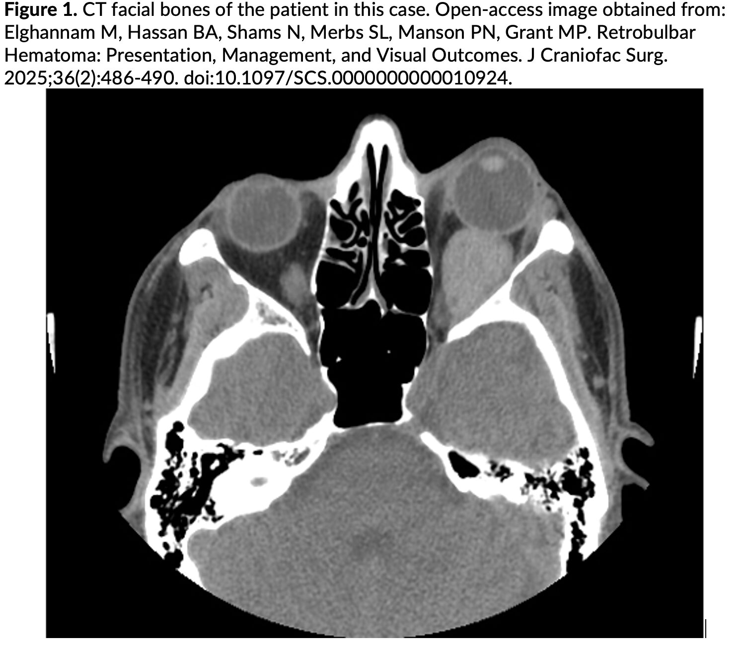
What is the diagnosis?
Answer: Retrobulbar Hematoma (RBH)
Background:
- RBH refers to bleeding into the space behind the eye, within the bony orbit
- Feared complication is orbital compartment syndrome (OCS), a vision-threatening ophthalmologic emergency1
- Orbital compartment is fixed; blood becomes trapped behind the eye, resulting in increased intraocular pressure, preventing arterial inflow and venous outflow, causing optic nerve ischemia2
Etiology:3
- Often results from blunt facial trauma, but may also occur postoperatively following maxillofacial/plastic/ophthalmologic surgery, or in rare cases, spontaneously
- Common mechanisms of injury in patients presenting to the ED with RBH
- Assault (36%)
- Motor Vehicle Collision (32%)
- Fall (28%)
- Other/unknown (4%)
Epidemiology:3
- Incidence
- <1% among all blunt facial trauma patients
- 6% of patients presenting to the ED with orbital fracture or orbital trauma
- 14% of permanent monocular blindness in patients presenting with orbital fracture or orbital trauma
- Up to 48% incidence of monocular blindness in patients with RBH
- More common in males (66%)
- More likely in patients on anticoagulation4
- Also more likely to have delayed RBH several days after initial orbital trauma
Clinical Presentation:3
- Eye pain
- Proptosis
- Chemosis
- Subconjunctival hemorrhage
- Periorbital edema
- Ophthalmoplegia
- Elevated IOP (>20 mmHg)
- Decreased visual acuity
- Relative afferent pupillary defect (RAPD)5,2
Evaluation:
- Standard trauma evaluation with primary and secondary survey6
- While the RBH may be the most obvious injury, do not forget to fully assess the patient for additional injuries
- Prompt ocular examination
- Worse visual outcomes for patients presenting >2 hours after injury, even with lateral canthotomy and cantholysis (LCC) intervention5
- Serial tonometry1,3
- Irreversible optic nerve ischemia can occur within 60-100 minutes based on animal data
- Q2H tonometry in the immediate post-injury period; spacing of checks after the first few hours without progression of symptoms or increasing IOP to be determined in conjunction with ophthalmology consultants7
- Intervention indicated for IOP > 408
- Contraindicated if evidence of globe rupture9
- Visual acuity testing10
- Extraocular movement testing for ophthalmoplegia
- Swinging flashlight test for relative afferent pupillary defect (RAPD)
- CT facial bones to assess for hematoma, associated fractures, etc.
- If clinical evidence of OCS, CT may be deferred until after emergent LCC2
- Emergent ophthalmology consultation
Diagnosis:
- OCS is a clinical diagnosis1
- Indicated by severe unilateral eye pain, proptosis, tight eyelids that are difficult to retract, ophthalmoplegia, monocular vision loss, RAPD, and elevated IOP10
- Presence and size of RBH and associated facial fractures identified on advanced imaging, usually CT of the facial bones1
Treatment:
- Reverse anticoagulation, as RBH is a vision-threatening bleed11
- Small RBH with normal serial tonometric exam and intact visual acuity may be managed medically3
- Adjunctive treatments to lower IOP include O2, IV steroids, acetazolamide, mannitol, ophthalmic beta-blockers, pain control
- Any increase in IOP or worsening visual acuity should be managed with LCC
- Emergent LCC for patients with increasing IOP on serial tonometry or signs/symptoms of orbital compartment syndrome (OCS) with optic nerve ischemia12
- Performing a LCC:10 (https://emdocs.net/wp-content/uploads/2014/09/OrbitalCompartmentSyndrome-Trevias-.pdf)
- Systemic analgesia and/or anxiolysis (e.g., fentanyl and midazolam, sub-dissociative ketamine 0.3 mg/kg IV)
- Inject local anesthetic (1% lidocaine with epinephrine) into the lateral canthus
- Clamp hemostat from lateral canthus to outer orbital rim; keep clamped for 30-60 seconds to crush tissue and optimize hemostasis
- Use iris scissors to cut from lateral canthus to outer orbital rim along the crushed tissue
- Grasp the lower lid with tissue forceps and expose the inferior crus of the lateral canthal tendon
- Cut the inferior crus of the lateral canthal tendon using iris scissors
- Recheck tonometry, pupils, and visual acuity testing
- If IOP does not improve, also cut the superior crus of the lateral canthal tendon using iris scissors
- Recheck tonometry, pupils, and visual acuity testing
- An alternative method is the vertical lid split procedure, where a full-thickness vertical 5 mm incision using iris scissors is made at the junction of the lateral 1/3 of the upper and lower eyelid.
- In patients for whom LCC or vertical lid split is insufficient, second-line treatment involves orbital decompression in the operating room, preferably within 6 hours of the trauma7
- Disposition: Admit for serial tonometry, visual acuity testing, and pain control3
Prognosis:
- Presentation/decompression within 2 hours of injury is associated with more favorable visual outcomes based on retrospective data,13 but larger studies have found that decompression within 6 hours of injury may be acceptable7
- Factors associated with improved outcomes14
- Prompt presentation/treatment
- Younger age
- Decreased severity/number of symptoms
- Absence of polytrauma
- Surgical decompression
- Favorable visual acuity on presentation15
Pearls:
- OCS is a vision-threatening emergency, often resulting from traumatic RBH.
- Signs of RBH with OCS include unilateral eye pain, proptosis, ophthalmoplegia, monocular vision loss, RAPD, and elevated IOP.
- CT can assist with the identification of RBH, but should not delay intervention if there is clinical evidence of OCS.
- Requires ophthalmology consultation for specialized care, but do not delay decompression for consultant arrival or transfer to a tertiary center.
- LCC are most effective when performed within 2 hours of injury.

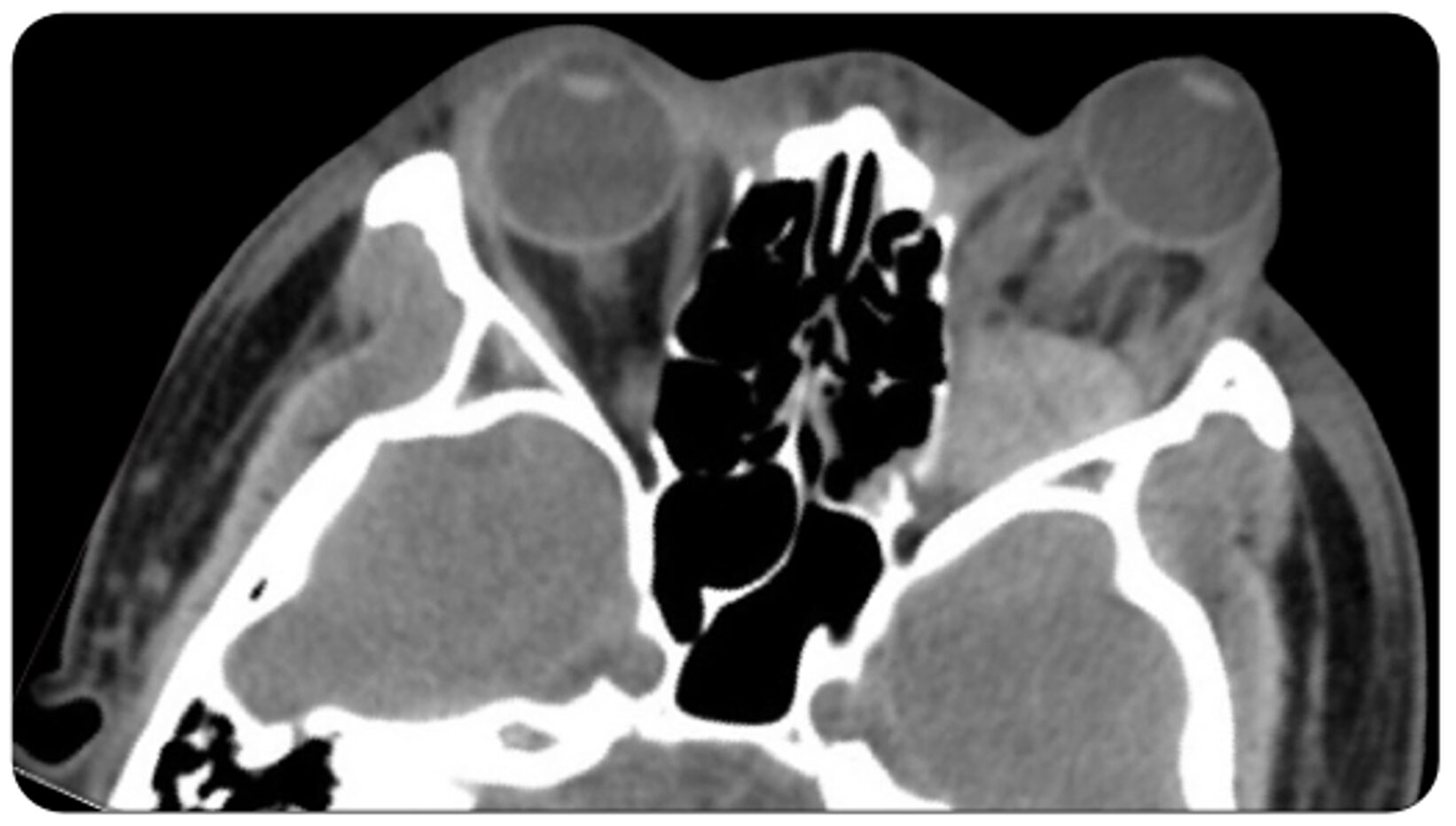
A 30-year-old man presents to the ED with severe left eye pain after being struck in the face with a line drive during a baseball game while coaching third base. On physical exam, his visual acuity is 20/30 on the right and hand waving on the left. A noncontrast computed tomography scan of the orbits is shown above. Which of the following is a physical exam finding often associated with this patient’s injury?
A) Herniation of the globe
B) Lateral gaze palsy
C) Severe proptosis
D) Teardrop-shaped pupil
Answer: C
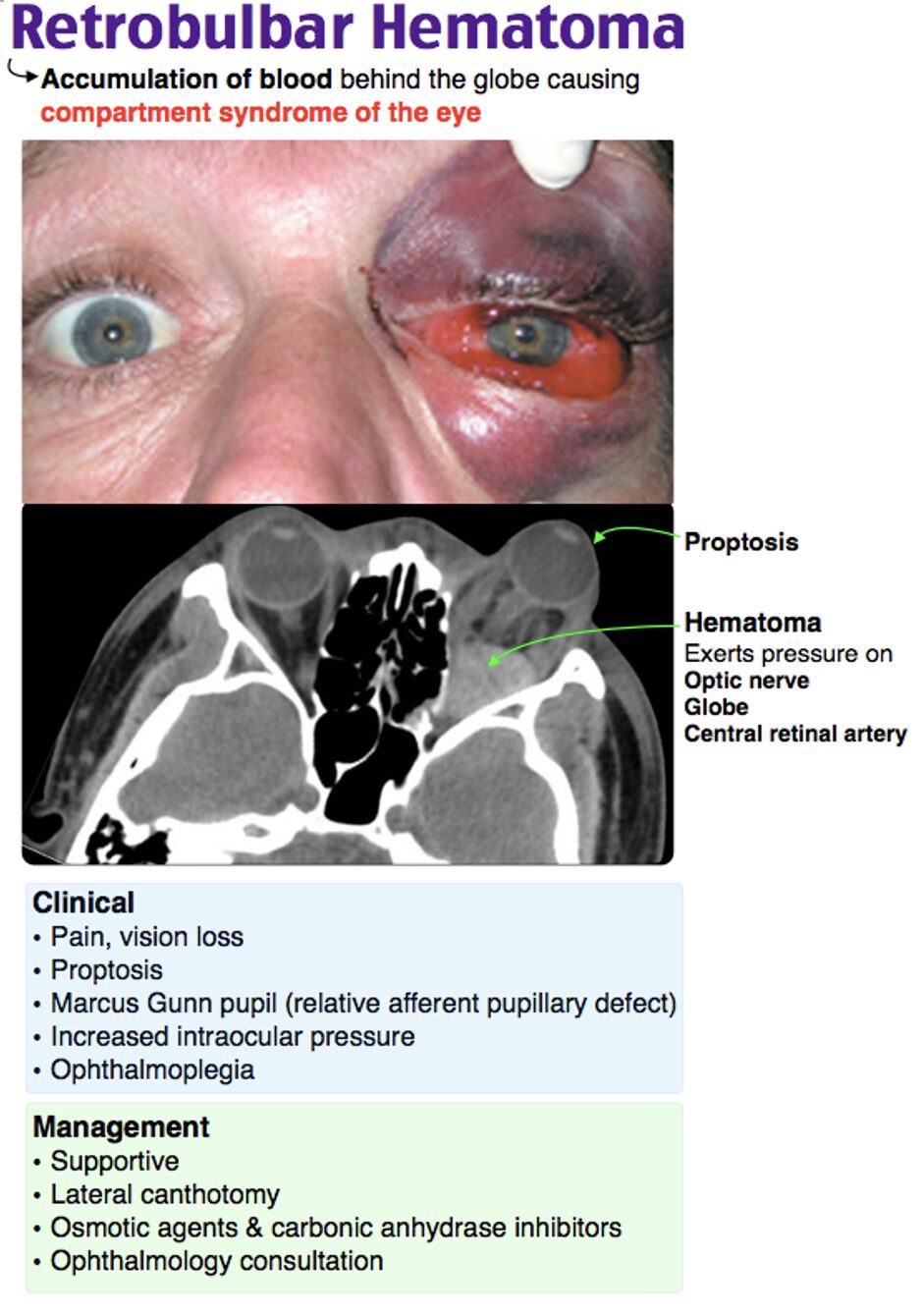
This patient has evidence of a retrobulbar hematoma. A retrobulbar hematoma is an accumulation of blood behind the globe that causes compartment syndrome of the eye by placing pressure on the optic nerve, the globe, and the central retinal artery. This is most commonly post-traumatic in etiology. Signs and symptoms of retrobulbar hematoma include pain, vision loss, proptosis, increased intraocular pressure, ophthalmoplegia or paralysis of the extraocular muscles, and an afferent pupillary defect.
Diagnosis is typically clinical, although a CT of the orbits may be necessary if the diagnosis is unclear. Management of retrobulbar hematoma is an emergent lateral canthotomy to relieve rapidly rising intraocular pressure, which can lead to irreversible vision loss within 90–120 minutes. Temporizing measures may be necessary depending upon available resources and so osmotic agents and carbonic anhydrase inhibitors may be used for this purpose. Ophthalmology should also be consulted immediately upon suspicion of this diagnosis.
Herniation of the globe (A) may be seen in severe orbital blowout fractures involving the orbital floor. This often results in traction on the optic nerve, which may lead to vision loss.
Lateral gaze palsy (B) is often seen with abducens nerve palsy, as the sixth cranial nerve is responsible for the lateral rectus muscle.
A teardrop-shaped pupil (D) is characteristically seen with globe rupture.
Further Reading:
Further FOAMed:
- https://emdocs.net/wp-content/uploads/2014/09/OrbitalCompartmentSyndrome-Trevias-.pdf
- https://emdocs.net/unlocking-common-ed-procedures-lateral-canthotomy-and-cantholysis/
- https://eyewiki.org/Orbital_Compartment_Syndrome
- https://wikem.org/wiki/Retrobulbar_hemorrhage
References:
- Ahmed A, Ameen A, Mahato RK. Retrobulbar hematoma in oral and maxillofacial trauma: an emergency requiring prompt recognition. Ann Med Surg. 2026;88(2):2172-2173. doi:10.1097/MS9.0000000000004646
- Peisah RI, Ostrowski K. Emergency management of orbital compartment syndrome: Lateral canthotomy and cantholysis case series. Australas Emerg Care. 2025;28(1):67-71. doi:10.1016/j.auec.2024.09.004
- Fattahi T, Brewer K, Retana A, Ogledzki M. Incidence of Retrobulbar Hemorrhage in the Emergency Department. J Oral Maxillofac Surg. 2014;72(12):2500-2502. doi:10.1016/j.joms.2014.06.457
- Berg BI, Flury E, Thieringer FM, et al. Retrobulbar haematoma in the era of anticoagulants. Injury. 2019;50(10):1641-1648. doi:10.1016/j.injury.2019.08.035
- Narjus-Sterba M, Puolakkainen T, Kokko L, Thorén H, Snäll J. Occurrence and outcomes of retrobulbar haematoma in 2149 orbital fracture patients. Oral Maxillofac Surg. 2025;29(1):134. doi:10.1007/s10006-025-01427-2
- Thim T, Krarup NHV, Grove EL, Rohde CV, Løfgren B. Initial assessment and treatment with the Airway, Breathing, Circulation, Disability, Exposure (ABCDE) approach. Int J Gen Med. 2012;5:117-121. doi:10.2147/IJGM.S28478
- Bourquard F, Foy JP, Hennocq Q, et al. Diagnosis and management of orbital compartment syndrome caused by retrobulbar hematoma following orbital fractures. J Cranio-Maxillo-fac Surg Off Publ Eur Assoc Cranio-Maxillo-fac Surg. 2026;54(3):104445. doi:10.1016/j.jcms.2026.104445
- Indarawis DW. Initial Evaluation and Management of Ophthalmic and Maxillofacial Trauma in Adults. Physician Assist Clin. 2025;10(4):611-623. doi:10.1016/j.cpha.2025.06.001
- Pelletier J, Koyfman A, Long B. High risk and low prevalence diseases: Open globe injury. Am J Emerg Med. Published online December 2022:S0735675722007410. doi:10.1016/j.ajem.2022.11.036
- Pelletier J. A Case of Painful Visual Loss- Managing Orbital Compartment Syndrome in the Emergency Department. Published online 2024. doi:10.21980/J8N35D
- Berg BI, Flury E, Thieringer FM, et al. Retrobulbar haematoma in the era of anticoagulants. Injury. 2019;50(10):1641-1648. doi:10.1016/j.injury.2019.08.035
- Sun MT, Chan WO, Selva D. Traumatic orbital compartment syndrome: Importance of the lateral canthotomy and cantholysis. Emerg Med Australas. 2014;26(3):274-278. doi:10.1111/1742-6723.12236
- Sun MT, Chan WO, Selva D. Traumatic orbital compartment syndrome: importance of the lateral canthotomy and cantholysis. Emerg Med Australas EMA. 2014;26(3):274-278. doi:10.1111/1742-6723.12236
- Christie B, Block L, Ma Y, Wick A, Afifi A. Retrobulbar hematoma: A systematic review of factors related to outcomes. J Plast Reconstr Aesthet Surg. 2018;71(2):155-161. doi:10.1016/j.bjps.2017.10.025
- Elghannam M, Hassan BA, Shams N, Merbs SL, Manson PN, Grant MP. Retrobulbar Hematoma: Presentation, Management, and Visual Outcomes. J Craniofac Surg. 2025;36(2):486-490. doi:10.1097/SCS.0000000000010924