Authors: Monica Yassa MD (Emergency Medicine PGY-4 Rutgers New Jersey Medical School, Newark, NJ), Ivan Ivanov DO (Faculty, Department of Emergency Medicine and Division of Medical Toxicology, Rutgers New Jersey Medical School, Newark, NJ), Anita Mudan MD (Faculty, Department of Emergency Medicine and Division of Medical Toxicology, Rutgers New Jersey Medical School, Newark, NJ) // Reviewed by Ann-Jeannette Geib, MD (Associate Professor of Emergency Medicine, Atrium Health Carolinas Medical Center and Wake Forest University School of Medicine, Charlotte, NC); Brit Long, MD (@long_brit)
Case:
A 56-year-old female with past medical history of hypertension, diabetes mellitus, cocaine use, and opioid use presents to the emergency department (ED) by emergency medical services (EMS) after being found altered, surrounded by emesis and drug paraphernalia. She received 2 mg intranasal naloxone by EMS with some improvement of mental status, but remains altered on arrival to the ED.
Vital signs
HR 73, BP 112/55, PO2 95% on 6L nasal cannula, RR 16, T 97.8F
On physical exam, she is ill-appearing and disoriented. Pupils are equal, 4mm, round, and reactive to light. She is protecting her airway. She opens her eyes to sternal rub and moves all four extremities in response to painful stimuli but is unable to follow commands.
The patient remains persistently altered despite receiving naloxone. CT of the head is obtained to evaluate for other possible causes of altered mental status. The CT read is notable for diffuse hypodensities of the cerebellar white matter with vasogenic edema and hypodensities of the globus pallida bilaterally.
Questions:
- What is CHANTER Syndrome?
- How does CHANTER Syndrome present clinically?
- What is the importance of recognizing CHANTER Syndrome?
Background:
- Cerebellar, Hippocampal, And basal Nuclei Transient Edema with Restricted diffusion (CHANTER) Syndrome is a clinical diagnosis supported by a distinct pattern of restricted diffusion on MRI.1
- It is often seen following an acute overdose, commonly of opioids and/or cocaine.1
- It is thought to be related to a combination of hypoxia and direct neurotoxic effects of opioids on the brain, which preferentially affect the cerebellum, hippocampus, and basal nuclei.1,2
- CHANTER involves distinct areas of the brain suggesting neurotoxins from certain drugs play a significant role. This distinguishes CHANTER from other differential diagnoses such as Hypoxic Ischemic Encephalopathy (HIE) or Heroin Associated Spongiform Leukoencephalopathy (HASL).
- The first case report of CHANTER syndrome was described in 2019 and since then, there have been several further case reports and series.1,3
- CHANTER syndrome is a relatively new diagnosis and is exceedingly rare, likely because it has been under-recognized and under-reported.
Clinical Presentation:1,3,4
Drug intoxication (usually opioids, or less commonly, cocaine) plus one of the following:
- Severe persistent headaches
- Persistent altered mental status longer than expected for the reported intoxication
- Minimal improvement in respiratory status after reversal with Naloxone
- Encephalopathy
- Seizures
- Unresponsiveness
- Apnea
- Posturing
Diagnosis:
- CT: May vary from no acute findings, hypodensities in the cerebellum (less often in the basal ganglia), or cerebellar edema +/- obstructive hydrocephalus. 3–5
- Diffusion-weighted MRI: symmetric diffusion restriction in bilateral gray matter of cerebellar cortex and hippocampi, asymmetric diffusion restriction in bilateral basal nuclei, with no white matter involvement. 1,3
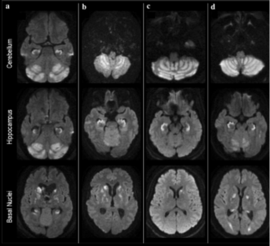
Figure 1 Diffusion weighted MRI of four patients diagnosed with CHANTER Syndrome (Jasne et al, 2019)
Management:1,3
- Symptomatic/supportive treatments have been described:
- Intubation for respiratory failure or airway protection
- Benzodiazepines for seizures
- Anti-emetics for nausea/vomiting
- Treatment of cerebral edema and increased intracranial pressure are key, after resuscitative measures are undertaken:
- Consultation with neurology +/- neurosurgery
- Hypertonic saline or mannitol
- Some require External Ventricular Drain (EVD) for Cerebrospinal Fluid (CSF) drainage
- Possible decompressive craniectomy
- Physical therapy / occupational therapy
Case Follow-up:
During the hospital course, the patient’s mental status rapidly declined, and she became completely obtunded, requiring intubation. Repeat CT head showed diffuse hypoattenuation in the bilateral cerebellum with mass effect on the fourth ventricle, causing moderate noncommunicating hydrocephalus. MRI shortly after as read by radiology showed diffusion restriction in the cerebellum, hippocampus, and basal ganglia concerning for CHANTER Syndrome secondary to opioid neurotoxicity versus bilateral posterior circulation infarction/ischemia secondary to cocaine abuse. Neurology and neurosurgery were consulted to see the patient. The patient received mannitol, was intubated for EVD placement, and eventually required a suboccipital decompressive craniectomy. After a prolonged hospital course, her mental status improved, and she was able to open her eyes and minimally move her extremities. She required a tracheostomy and PEG tube placement and was ultimately discharged to a long-term acute care facility.
Clinical Pearls:
-
- Patterns with large areas of diffusion restriction are often thought to have irreversible damage and a poor prognosis. Early recognition of CHANTER syndrome with aggressive medical and possible surgical management can result in improved prognosis compared to other etiologies with similar clinical/radiographic presentations.3–5
- CHANTER Syndrome is likely underdiagnosed in patients who have more subtle symptoms and no clear indication to obtain a CT of the head.
- A high index of suspicion and early consultation can position patients to receive potentially lifesaving interventions.
- CHANTER Syndrome is a progressive condition that can present anywhere along a spectrum between severe, persistent headaches and unresponsiveness or posturing. Thus, findings on CT imaging may be variable. The pathophysiology of the progression of the disease is still unclear.
- Other differentials to consider (details in the Table)
- Heroin Associated Spongiform Leukoencephalopathy (HASL)
- Posterior Reversible Encephalopathy Syndrome (PRES)
- Hypoxic Ischemic Encephalopathy (HIE) / Anoxic Brain Injury
- Opioid Amnestic Syndrome
- Other co-intoxicant or substance contaminants
- Acute Ischemic Stroke
- Intracranial hemorrhage (ie, epidural hematoma, subdural hematoma, intraparenchymal hemorrhage, subarachnoid hemorrhage)
- Traumatic brain injury (TBI)
- Cerebral artery dissection
- Cerebral venous sinus thrombosis
- CNS infection (ie, meningitis, encephalitis)
DISEASE PROCESS RISK FACTORS CLINICAL FINDINGS CT FINDINGS MRI FINDINGS
CHANTER1,3–5(Cerebellar, Hippocampal, And basal Nuclei Transient Edema with Restricted diffusion)*Opioid > amphetamine exposure *Headache *Confusion
*Altered mental status
*Unresponsive/comatose
*Respiratory depression
*Signs of ↑ ICP
+/- cerebellar edema +/- obstructive hydrocephalus
*Symmetric diffusion restriction in gray matter of cerebellar cortex and hippocampi *Asymmetric diffusion restriction in bilateral basal nuclei, with
*No white matter involvement
HASL1,5–7 (Heroin Associated Spongiform Leukoencephalopathy)
*Inhalation of opioid vapors (heroin > fentanyl) *Cerebellar neurologic abnormalities *Hyperreflexia
*Spastic paresis
*Myoclonic jerks
*Cognitive dysfunction
*Posturing (severe stages)
+/- white matter hypodensity *Symmetric T2/FLAIR white matterhyperintensities in cerebellum, posterior cerebrum, corpus callosum, brainstem *Sparing of cortex and basal ganglia
PRES1,10 (Posterior Reversible Encephalopathy Syndrome)
*Acute hypertension *Immunosuppression
*Autoimmune disease
*Renal Failure
*Eclampsia
*Seizures *Headache
*Confusion
*Visual disturbances
*Altered mental status
+/- vasogenic edema in parieto-occipital or fronto-temporal regions +/- intracranial hemorrhage
*Subcortical vasogenic edema T2/FLAIR hyperintensity *Commonly involves parieto-occipital or fronto-temporal regions *Primarily involves white matter
HIE1,3,5,8,9 (Hypoxic Ischemic Encephalopathy Syndrome)
*Cardiac arrest *Prolonged hypoxia
*Near drowning
*Asphyxiation
*Not associated with drug exposure
*Unresponsive/comatose *Absent brainstem reflexes
*Multi-organ injury
Loss of gray-white matter differentiation *Restricted diffusion in cortex(specially watershedzones) OAS5,12,13 (Opioid Amnestic Syndrome)
*Opioid use (fentanyl > heroin) *Sudden onset isolated anterograde amnesia *Normal neuro exam
Typically normal *Bilateral hippocampalrestricted diffusion (DWI hyperintensity), T2/FLAIR hippocampal hyperintensity Acute Ischemic Stroke11 *Cardiovascular disease *Cardiac dysrhythmia
*Hyperlipidemia
*Hypercoagulable state
*Sudden onset focal neuro deficit Typically normal =/- subtle loss of gray-white differentiation
*Restricted diffusion (DWI hyperintensity) in a defined vascular territory ICH14 (Intracerebral Hemorrhage)
*Hypertension *Anticoagulation use
*Head trauma
*Focal neuro deficit *Headache
*Nausea/vomiting
*Altered mental status
*Unresponsive/comatose
Hyperdense lesion with surrounding edema Appearance varies by age of blood products
References:
- Jasne AS, Alsherbini KH, Smith MS, Pandhi A, Vagal A, Kanter D. Cerebellar Hippocampal and Basal Nuclei Transient Edema with Restricted diffusion (CHANTER) Syndrome. Neurocrit Care. 2019;31(2):288-296. doi:10.1007/s12028-018-00666-4
- Oehmichen M, Meißner C, Reiter A, Birkholz M. Neuropathology in non-human immunodeficiency virus-infected drug addicts: hypoxic brain damage after chronic intravenous drug abuse. Acta Neuropathol (Berl). 1996;91(6):642-646. doi:10.1007/s004010050478
- Pandit R, Hamki O, Gaddamanugu S. Cerebellar, Hippocampal, and Basal Nuclei Transient Edema With Restricted Diffusion (CHANTER) Syndrome: Radiologic Features and Findings. Korean J Radiol. 2024;25(3):314. doi:10.3348/kjr.2023.1142
- Suthar PP, Hughes K, Mafraji M, Akyuz M, Jhaveri M, Dua SG. Case 324: CHANTER Syndrome. Radiology. 2024;311(1):e222748. doi:10.1148/radiol.222748
- Mallikarjun KS, Parsons MS, Nigogosyan Z, Goyal MS, Eldaya RW. Neuroimaging Findings in CHANTER Syndrome: A Case Series. AJNR Am J Neuroradiol. 2022;43(8):1136-1141. doi:10.3174/ajnr.A7569
- Keogh CF, Andrews GT, Spacey SD, Forkheim KE, Graeb DA. Neuroimaging Features of Heroin Inhalation Toxicity: “Chasing the Dragon.” Am J Roentgenol. 2003;180(3):847-850. doi:10.2214/ajr.180.3.1800847
- Kriegstein AR, Shungu DC, Millar WS, et al. Leukoencephalopathy and raised brain lactate from heroin vapor inhalation (“chasing the dragon”). Neurology. 1999;53(8):1765-1765. doi:10.1212/WNL.53.8.1765
- Gutierrez LG, Rovira A, Portela LAP, Leite C da C, Lucato LT. CT and MR in non-neonatal hypoxic-ischemic encephalopathy: radiological findings with pathophysiological correlations. Neuroradiology. 2010;52(11):949-976. doi:10.1007/s00234-010-0728-z
- Lang M, Kenda M, Scheel M, et al. Radiological signs of hypoxic-ischaemic encephalopathy on head computed tomography for prediction of poor functional outcome after cardiac arrest – a prospective observational cohort study. Resuscitation. 2025;214:110675. doi:10.1016/j.resuscitation.2025.110675
- Fugate JE, Rabinstein AA. Posterior reversible encephalopathy syndrome: clinical and radiological manifestations, pathophysiology, and outstanding questions. Lancet Neurol. 2015;14(9):914-925. doi:10.1016/S1474-4422(15)00111-8
- Czap AL, Sheth SA. Overview of Imaging Modalities in Stroke. Neurology. 2021;97(20 Suppl 2):S42-S51. doi:10.1212/WNL.0000000000012794
- Barash JA, Whitledge J, Watson CJ, et al. Opioid-associated amnestic syndrome: Description of the syndrome and validation of a proposed definition. J Neurol Sci. 2020;417:117048. doi:10.1016/j.jns.2020.117048
- Barash JA, Ganetsky M, Boyle KL, et al. Acute Amnestic Syndrome Associated with Fentanyl Overdose. N Engl J Med. 2018;378(12):1157-1158. doi:10.1056/NEJMc1716355
- Sheth KN. Spontaneous Intracerebral Hemorrhage. Ropper AH, ed. N Engl J Med. 2022;387(17):1589-1596. doi:10.1056/NEJMra2201449