Author: Erica Simon, DO, MHA (@E_M_Simon, EMS Fellow, USAF) // Edited by: Alex Koyfman, MD (@EMHighAK, EM Attending Physician, UT Southwestern Medical Center / Parkland Memorial Hospital) and Brit Long, MD (@long_brit)
A 55-year-old African American female presents to the emergency department for one month of progressively worsening dyspnea associated with bilateral lower extremity swelling. The patient reports occasional aching, left-sided chest pain, with the most recent episode occurring one hour prior to arrival, lasting 10 minutes, and improved with rest. The woman denies that her chest pain is associated nausea and diaphoresis. She denies orthopnea and paroxysmal nocturnal dyspnea. The patient denies cardiac risk factors, recent surgeries, hospitalizations, and a personal/familial history of PE/DVT. Previous medical history is significant for sarcoidosis (diagnosis two years prior by incidental finding on CXR; asymptomatic).
Initial VS: BP 147/92, HR 101, RR 14, T 99.8F, SpO2 97% on room air.
Physical examination findings:
CV: tachycardia, no murmurs, rubs, or gallops
Pulm: fine bibasilar inspiratory crackles
Abdomen: prominent hepatic border palpable 4cm below the right costal margin; hepato-jugular reflex present; + abdominal fluid wave
MSK: + 3 pitting edema bilaterally
This case depicts a patient with isolated right-sided heart failure. As you reviewed the HPI, vital signs, and examination, was the patient’s history of asymptomatic sarcoidosis meaningful in the development of your evaluation and treatment plan? If not, this review will offer insight as to why this patient population requires special consideration.
In this piece, we’ll briefly highlight facts regarding the epidemiology, pathophysiology, and diagnosis of sarcoidosis. We’ll then visit common emergency department complaints as applied to a patient with previously diagnosed sarcoidosis, offering pearls regarding disease manifestations that should be on your radar.
Note: Lists of differential diagnoses detailed below are not all encompassing, but are presented as examples to inspire critical thinking.
Epidemiology
Sarcoidosis displays a worldwide distribution, with the highest annual incidence reported in northern European countries (e.g. Sweden: 64 per 100,000 individuals with radiographic evidence of disease).1 Occurring most frequently amongst females 20-40 years of age, population studies estimate the annual U.S. incidence of sarcoidosis as 46 per 100,000 inhabitants.1,2 Amongst this U.S. group, African Americans are disproportionately affected (35 new cases/100,000 population).2 As compared to persons of differing races, sarcoidosis in African Americans is more likely to be chronic, involve multiple organs, and lead to death.3
Pathophysiology
Sarcoidosis is an inflammatory disorder frequently characterized by multi-organ involvement. Today, the etiology of sarcoidosis is unknown. Experts hypothesize a role for genetics (HLA groups) and environmental exposures, however, scientific research has failed to demonstrate a correlation.1,4 Sarcoidosis is thought to originate in lung tissue where the activation of pulmonary macrophages results in an inflammatory cascade, recruiting CD4+ T cells which interact with antigen-presenting cells to form/maintain non-caseating granulomas.1,5 The mechanism of dissemination to additional organ systems is poorly understood.1
Immunologic studies have demonstrated that sarcoidosis is a disease characterized by reduced peripheral cell-mediated immunity (peripheral lymphopenia and cutaneous anergy common1,6), and increased humoral immunity (polyclonal gammopathy and increased complement levels1,7), explaining the chronic inflammatory state and role for corticosteroid therapy.1
Sarcoidosis Diagnosis and Treatment
The diagnosis of sarcoidosis is one of exclusion. Nearly 50% of patients are diagnosed by incidental finding on chest radiograph (hilar adenopathy) and subsequent biopsy. The majority of patients with sarcoidosis are asymptomatic, do not require treatment, and have spontaneous remission within two years of diagnosis.5 The goal in treating symptomatic patients (demonstrating objective findings of organ dysfunction (PFTs, renal function tests, LFTs, etc.) is to preserve remaining organ function.
How does this affect your patient evaluation and treatment? The patient will often possess insight regarding his/her disease and will frequently be able to provide details regarding personal disease manifestations, organ dysfunction, required treatment(s), and methods for disease monitoring.
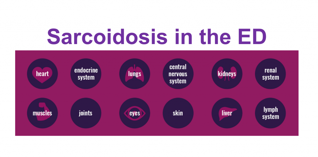
Manifestations for Emergency Physician (EP) Consideration
Let’s move on to the high-yield aspect of our review: chief complaints and the sarcoidosis patient:
Sarcoidosis & Altered Mental Status:
- Neurosarcoidosis (up to 25% of patients with sarcoidosis;5 more often occurring in females8):
- Psychiatric disturbance, altered level-of-consciousness, or seizure (seizures occur in up to 1/5th of patients with sarcoidosis9) = result of granulomatous lesions: aseptic meningitis, obstructing hydrocephalus, or CNS parenchymal disease.10,11
- Hypercalcemia (hallmark of sarcoidosis present in 2-63% of patients12): granulomatous macrophages convert 25-hydroxy-vitamin D to active 1,25-hydroxy-vitamin D:1,12
Evaluation:1,5,12
- Utilize the H&P to direct evaluation and imaging.
- Neurosarcoidosis differential diagnosis: TB, coccidiomycosis, parasitic infection, lymphoma.10 CT/MRI may support the diagnosis/allow for an analysis of lesion progression (if previous imaging) or hydrocephalus, but are non-specific.10 CSF sensitive and specific: lymphocytosis and elevated protein (occasionally reduced glucose). Histologic examination = gold standard.
- Hypercalcemia: serum ionized calcium, renal function testing,13 +/- UA (hypercalciuria), EKG.
Treatment:5,12
- Consult as appropriate.
- Address the underlying etiology: recommended that patients with acute neurologic symptoms and hypercalcemia receive 40-80 mg PO prednisone QD for 8-12 weeks with tapering of the dose to10 mg QOD over 8 -12 months.5,12
- Hypercalcemia: IVF therapy (forced diuresis).12
Sarcoidosis & *Focal Neurologic Deficit
*Futher discussion of CN II and ocular pathology to follow.
- Neurosarcoidosis manifestations:
- Cranial neuropathies = most frequent manifestation.1,7 Patients may present with CN VII palsy (most common), CN II involvement (second most common: presenting with papilledema and/or decreased visual acuity), and less frequently palsies of CNs III, IV, and VI, and CN VIII deficit.1,9,10
- Rare presentations: spinal cord lesions and their respective neurologic findings, peripheral neuropathy, and mononeuritis multiplex.1,12
Evaluation:1,5,12
- Utilize the H&P to direct evaluation and imaging.
- Neurosarcoidosis evaluation as above (i.e. CT/MRI, CSF, histological examination as appropriate). Additional differential diagnoses for consideration: cavernous venous sinus thrombosis, Bell’s palsy, stroke.
Treatment:5,12
- Consult.
- Patients with acute symptoms should receive 40-80 mg PO prednisone QD for 8-12 weeks with tapering of the dose to10 mg QOD over 8 -12 months.5,12
Sarcoidosis & Eye Complaints
- Ocular Involvement (occurs in 11-83% of patients14):
- Lacrimal duct infiltration
- Orbital granulomatous disease = proptosis or exopthalmosis1
- Uveitis (most common presentation1,14); patients may report floaters and decreased visual acuity.1
- Retinal periphlebitis (posterior ocular segment); patients may experience flashes, floaters, painful loss of vision, blurred vision, alterations in color vision, and scotomata.14
- Optic nerve involvement: Optic neuritis; patients often present with painful loss of vision and/or notable color deficit.15
Evaluation:1,5,12
- Visual acuity, slit lamp examination, AND dilated eye examination (posterior segment involvement in 25% of patients with ocular sarcoidosis.1,9 Posterior pathology often requires advanced imaging: e.g. fluorescein angiography for posterior uveitis9,12). Differential diagnosis: infectious etiologies (ocular syphilis, CMV retinitis, HSV retinitis, TB uveitis, etc.), retinal detachment, etc.).
Treatment:5,12
- Consult ophthalmology for a complete examination and a discussion of therapy. Specifics cited in the literature:
- Iridiocyclitis: corticosteroid gtt + intraocular steroid injection.5
- Posterior uveitis: systemic corticosteroids.5
- Optic neuritis: often recommend a prolonged steroid taper in sarcoidosis patients given the refractory nature of the condition.15
Sarcoidosis & Upper Respiratory Tract Complaints
Involvement is less common, however, patients may present for recurrent sinus pain, rhinorrhea, epistaxis, and/or frontal headache.5,12
Evaluation:
- Perform a thorough H&P to direct laboratory evaluation and imaging.
Treatment:
- Refer to specialist as appropriate: requires biopsy to differentiate from other granulomatous diseases (actinomycosis, TB, and Wegener’s granulomatosis).1
Sarcoidosis & Airway/Oropharyngeal Complaints
- Salivary Glands (occurs in up to 70% of patients with sarcoidosis1):
- Unilateral or bilateral painless enlargement of the parotid gland (s) most common.1
- Chronic: may present with facial nerve weakness secondary to neural ischemia from perivascular lymphocytic infiltration.
- Pharynx involvement:
- Patients more frequently report dysphagia found to occur secondary to hilar lymphadenopathy.1
- Hypopharynx/Larynx involvement (1-5% of patients with sarcoidosis16,17):
- The epiglottis is the most commonly involved structure in the larynx.16 Patients may present with hoarseness, stridor, or cough.
- Unilateral or bilateral painless enlargement of the parotid gland (s) most common.1
Evaluation:1,5,12
- Assess airway and breathing; intervene as appropriate.
- Utilize the H&P to direct evaluation and imaging.
- Salivary gland enlargement: Differential diagnosis: infection (mumps – bilateral parotitis), abscess, malignancy, sialadenitis.
- Phaynx (pharyngeal dysphagia): Differential diagnosis: CVA (full neuro exam) vs. compression secondary to hilar lymphadenopathy (Consider CXR).
- Hypopharynx/Larynx: differential diagnosis: angioedema, anaphylaxis, foreign body, epiglottis, bacterial tracheitis.
Treatment:5,12,17,18
- Airway intervention if unstable. Consultation is optimal: laryngoscopic evaluation warranted when concern for airway involvement. Indication for IV steroid therapy.
Sarcoidosis & Chest Pain/Palpitations or Dyspnea
Cardiac involvement (5% of patients symptomatic1): infiltrative process: Depending on the etiology, patients may report palpitations, dizziness, syncope, dyspnea, and/or chest pain:12
- Conduction abnormalities: Infiltrative process may obliterate conduction pathways or granulomas act as arrythmogenic foci:1 Complete heart block most common initial presentation.19 However, patients may present in VT or Vfib arrest.
- Structural remodeling: ventricular aneurysms, and mitral valve dysfunction may occur.20
- Myocarditis syndrome: fevers, malaise, chest pain, dyspnea, arthralgias, myalgias secondary to chronic inflammation.1
- Pericardial effusion: secondary to chronic inflammatory state; may occur with tamponade causing cardiovascular compromise.12
- Cor pulmonale: frequently occurs secondary to pulmonary fibrosis resulting in prolonged pulmonary hypertension.
- Acute valvular regurgitation +/- heart failure: papillary muscle rupture secondary to granulomatous infiltration.
Evaluation:5,12
- EKG, CXR, troponin, BNP (as dictated by H&P), BMP +/- bedside echo.
- Differential diagnosis: coronary artery dissection, infectious myocarditis, myocardial infarction, electrolyte disturbance (arrhythmia).
Treatment:5,12
- Stabilization as appropriate (e.g. acute valve rupture: nitrates, etc.)
- Consultation as appropriate (cardiology, pulmonology).
- Corticosteroids depress conduction problems, suppress dysrhythmias and reduce the myocardial granuloma load.21
- Transplant is an option, but recurrent cardiac sarcoidosis has been reported within 6-19 months.22
Pulmonary involvement occurs in > 90% of patients with sarcoidosis (50% receive the diagnosis by incidental finding on CXR):5 patients often report dry cough, dyspnea, pleuritic chest pain, and rarely hemoptysis.1
Evaluation:5,12
- EKG, CXR, troponin, BNP (as dictated by H&P), CT chest (pulmonary angiography vs. non-contrast).
- Differential diagnosis: myocardial infarction, myocarditis, pericarditis, heart failure, pneumothorax, pneumomediastinum, malignancy, pulmonary embolism, pneumonia (fungal/bacterial/viral), restrictive lung disease (COPD, asthma), GERD (persistent dry cough).
Treatment:5,12
- Stabilization as appropriate.
- Consultation as appropriate (pulmonology): PFT monitoring (restrictive pattern), monitoring of pulmonary pressures, treatment of pulmonary hypertension.
- Steroids commonly advised for symptomatic patients or those in whom imaging is static or worsens over a three-month period.5
Sarcoidosis & Abdominal Pain or GI Bleed
Granulomatous hepatic infiltration is common, (20% of patients display hepatomegaly on exam1). Patients may present with abdominal pain and pruritus.12 Feared complications occurring secondary to hepatic involvement:
- Variceal bleed (portal hypertension), intrahepatic cholestasis, hepatic failure, hepatic encephalopathy, primary biliary cirrhosis, primary sclerosing cholangitis.12,23
- Note: fever is associated with hepatic sarcoidosis.1,12
Evaluation:5,12
- LFTs, coagulation panel, +/- RUQUS (symptomatic patients: elevation of alkaline phosphatase and transaminases common12). Differential diagnosis: malignancy, hepatic abscess, cholecystitis, choledocolithiasis, hepatic steatosis.
Treatment:5,12
- Stabilization as appropriate (e.g. gastric variceal bleed secondary to portal hypertension: type and screen vs. cross, protonoix, octreotide, etc.)
- Consultation as appropriate (GI).
Splenic involvement, as demonstrated by splenomegaly on examination, is present in 5-14% of sarcoidosis patients.12 Individuals frequently report fever, abdominal pain and malaise.
- Hyperslenism may rarely occur.12
Evaluation:5,12
- CBC with peripheral smear +/- heterophile antibody test (based upon H&P). Differential diagnosis: malignancy, protozoal infection (malaria), mononucleosis.
Treatment:5,12
- Consultation as appropriate – splenomegaly generally responds to steroid therapy.1
Note: Rare involvement of the esophagus, stomach (most common1), pancreas, small bowl, appendix, colon, and peritoneum has been reported. May present as surgical emergencies: ileus, hemorrhage (granulomatous erosion into vasculature). Requires referral for biopsy (differentiation from Crohn’s disease).12
Sarcoidosis & Flank Pain
- Nephrolithiasis, nephrocalcinosis, interstitial calcium deposits, renal failure: occur secondary to hypercalcemia and renal granulomatous infiltration.
Evaluation:1,5,12
- Utilize the H&P to direct evaluation and imaging (serum ionized calcium, renal function testing,13 +/- UA, EKG; non-contrast CT abdomen pelvis (nephrolithiasis), vs. ultrasound, etc). Differential diagnosis: malignancy (renal cell carcinoma).12
Treatment:5,12
- Consult as appropriate.
- IVF therapy (forced diuresis)12
- Recommended that all hypercalcemic patients receive 40-80 mg PO prednisone QD for 8-12 weeks with tapering of the dose to10 mg QOD over 8 -12 months (controlled granulomatous formation of calcitriol (1,25-hydroxy-vitamin D).5
Sarcoidosis & Cutaneous Complaints
Patients with sarcodosis may suffer from cutaneous lesions (24.2%).12
- Erythema nodosum: tender subcutaneous nodule often seen on the anterior aspect of the tibial surface; most common skin finding. Demonstrates a female predominance.24
- Lupus pernio: classic sarcoid-specific skin finding: symmetric, red, or violaceous plaques localized to the nose, cheeks, ears, or digits. Lesions may erode into underlying cartilage and bone making them severely disfiguring.5,12
Numerous other skin manifestations may be found: papules, nodules, and maculopapular lesions.12
Evaluation:5,12
- Dictated by the H&P.
- Differential diagnosis for new onset lesions: TB, Group B streptococcal infection, fungal infections, coccidiomycosis, contact dermatitis, cutaneous manifestation associated with inflammatory bowel disease, etc.12
Treatment:5,12
- Consultation/referral as appropriate. Biopsy may be required to determine etiology. Corticosteroid therapy recommended for Lupus pernio.5
Sarcoidosis & Musculoskeletal Complaints
Individuals with sarcoidosis often suffer from acute or chronic arthritis (14-38%) and arthralgias (70%).12,24
- Acute arthritis: self-resolving; mono- or poly-articular; usually involving the ankles or knees and is associated with erythema nodosum.12
- Chronic arthritis: variant occurring most often in African American individuals with cutaneous sarcoidosis.12
Cystic bone lesions are reported in 1-13% of patients with sarcoidosis (majority localized to the bones of the hands and feet).12,24
The chronic inflammatory nature of sarcoidosis may also manifest as progressive myopathy and myositis.12
Evaluation:5,12
- Dictated by the H&P (e.g. myositis: BMP, CK, rapid influenza, etc.).
- Differential diagnosis: bone lesions: malignancy, myositis/myopathy: viral myositis, medication induced myositis (statins), autoimmune pathology (polymyositis, etc.).
Treatment:5,12
- Supportive care.
- Consultation/referral as appropriate. Corticosteroid therapy recommended in the case of symptomatic bone lesions.5
Sarcoidosis & Endocrine Abnormalities (Variable Manifestations)
Thyroid infiltration, parathyroid infiltration, and adrenal infiltration are uncommon.1 Incidental nodules or masses may be identified on imaging. Thyroid involvement (5% of sarcoidosis patients at autopsy1) may present as thyroiditis or in exceedingly rare cases, refractory thyrotoxicosis.25
Evaluation:12
- Utilize the H&P to direct evaluation and imaging (e.g. thyroid function tests, electrolytes, ionized calcium level, palpable thyroid mass: ultrasound, etc.)
Treatment:5,12
- Consult as appropriate: asymptomatic patients may not require treatment.5 Case studies of thyrotoxicosis secondary to granulomatous invasion of the thyroid have demonstrated improvement following the admission of systemic corticosteroids.13
Note: Neurosarcoidosis may also cause a variety of hypothalamic pituitary lesions with clinical symptoms depend upon the neurologic site of involvement: diabetes insipidus, amenorrhea-galactorrhea, syndrome of inappropriate antidiuretic hormone, adrenal insufficiency, secondary hypothyroidism.12 Evaluation and treatment as for neurosarcoidosis above.
Sarcoidosis & Hematopoietic Abnormalities
Bone marrow infiltration may occur in the patient with sarcoidosis. Leukopenia, lymphopenia, anemia, and thrombocytopenia may be evidenced on laboratory studies,1,12 however, individuals are rarely symptomatic.
Evaluation:5,12
- Dictated by the H&P (question specifically regarding B symptoms: CBC, peripheral smear, reticulocyte count).
- Differential diagnosis: reticuloendothelial malignancy.
Treatment:5,12
- Consultation/referral as appropriate: may require bone marrow biopsy.
Sarcoidosis & The Kitchen Sink
The following presentations are exceedingly rare, but documented, manifestations of sarcoidosis:
Sarcoidosis of the breast: may present as a mass lesion => biopsy to exclude malignancy.12
Sarcoidosis of the GU tract: granulomatous infiltration of the testis, epididymis, and prostrate have been documented in African American males (presentations: testicular pain and rectal pain) => rule out malignancy.12 Sarcoidosis may also affect the female reproductive tract (ovaries, fallopian tubes, uterus, and vulva) (presentations: abdominal pain, vulvar mass, etc.).12
The More You Know
Let’s take a minute to talk fever in the sarcoidosis patient:
Sarcoidosis is a chronic inflammatory disease, frequently associated with fever (as discussed previously). Be on your guard and review your differential diagnoses: a vital sign abnormality should not be attributed to the sarcoidosis diagnosis without careful consideration and appropriate evaluation.
Speaking the language:
When discussing a case with consultants or reading the literature, you may come across a number of eponyms. FYSA:
Heerfordt syndrome: anterior uveitis, parotiditis, fevers, and facial nerve palsy.5
Löfgren’s syndrome: arthritis, erythema nodosum, bilateral hilar adenopathy (occurs in 9-43% of patients).5
Mikulicz’s syndrome: bilateral lacrimal and salivary gland swelling/infiltration.1
TASS syndrome: thyroiditis, Addison’s disease, Sjogren’s syndrome, and sarcoidosis.1
Back to our case:
Our patient requires hospitalization with cardiology and pulmonology consults. An echo, assessment of pulmonary artery pressures, assessment of PFTs (crackles on exam indicating potential for fibrosis), a BAL, and a non-contrast CT chest are all reasonable diagnostic measures to evaluate/address the underlying etiology and initiate treatment (presumed pulmonary artery hypertension secondary to pulmonary sarcoidosis: PDE inhibitors, diuretics, steroids, etc.).
Key Pearls
- Sarcoidosis patients are complicated – be aware of the numerous disease manifestations that may result in morbidity and mortality.
- Fever may be a characteristic of several conditions resulting from granuloumatous organ invasion, but it requires vigilance to perform an appropriate evaluation for alternative underlying etiologies.
- Consult early. If your patient is suffering from a condition that might be caused by or complicated by his/her underlying sarcoidosis => call the experts.
References / Further Reading:
- Pollack C, Jorden R. Recognition and management of sarcoidosis in the emergency department. JEM. 1993; 297-308.
- Tybicki B, Major M, Popovich J, Maliarik M, Iannuzzi M. Racial differences in sarcoidosis incidence: a 5-year study in a health maintenance organization. Am J Epidemiol. 1997; 145:234-241.
- Israel H, Karlin P, Menduke H, DeLisster O. Factors affecting outcome of sarcoidosis. Influence of race, extrathoracic involvement, and initial radiologic lung lesions. Ann N Y Acad Sci. 1986; 465:609-618.
- American Thoracic Society. Sarcoidosis. Available from: https://www.thoracic.org/patients/patient-resources/breathing-in-america/resources/chapter-21-sarcoidosis.pdf
- Qawi I. Sarcoidosis. In Ferri’s Clinical Advisor 2018. Philadelphia, Elsevier. 2018; 1137-1138.e2.
- Bascom R, Johns C. The natural history and management of sarcoidosis. Adv Intern Med. 1986; 31:213-241.
- Lazarus A. Sarcoidosis. Disease-A-Month. 1990; 36:471-535.
- Stern B, Corbett J. Neuro-opthalmologic manifestations of sarcoidosis. Curr Treat Options Neurol. 2007; 9:63-71.
- Ellison E, Canalis R. Sarcoidosis of the head and neck. Clin Dermatol. 1986; 4:136-142.
- Stern B, Krumholz A, Johns C, Scott P, Nissm J. Sarcoidosis and its neurological manifestations. Arch Neurol. 1985; 42: 909-917.
- Heck A, Phillips L. Sarcoidosis and the nervous system. Neurol Clin. 1989; 7:641-654
- Holmes J. Lazarus A. Sarcoidosis: Extrathoracic manifestations. Disease-A-Month. 2009; 55 (11):675-692.
- Sharma O. Renal sarcoidosis and hypercalcemia. Cur Respir J Monogr. 2005; 10:220-232.
- Abu El-Asrar A, Herbort C, Tabbara K. Differential diagnosis of retinal vasculitis. Middle Ease Afr J Opthalmol. 2009; 16(4): 202-218.
- Pula J, MacDonald C. Current options for the treatment of optic neuritis. Clin Ophthalmol. 2012; 6:1211-1223.
- Gold R, Sager E. Oral sarcoidosis: review of the literature. J Oral Surg. 1976; 34:237-244.
- Sims H, Thakkar K. Airway involvement and obstruction from granulomas in African
- Devine K. Sarcoidosis and sarcoidosis of the larynx. Laryngoscope. 1965; 75:533-569.
- Flora G, Sharma O. Myocardial sarcoidosis: a review. Sarcoidosis. 1989. 6:97-106.
- Oakley C. Cardiac sarcoidosis. Thorax. 1989; 44:371-372.
- Swanton R. Sarcoidosis of the heart. Eur Heart J. 1989; 9(Suppl G): 169-174.
- Milman N, Anderson C, Mortensen S, et al. Cardiac sarcoidosis and heart transplantation. Lancet 1990; 336: 1579.
- Keeffe E. Sarcoidosis and primary biliary cirrhosis: literature review and illustrative case. Am J Med. 1987; 83:977-980.
- Judson M. Extrapulmonary sarcoidosis. Am J Respir Crit Care Med. 2001;164:1885-1889.
- Yanamandra U, Kotwal N, Menon A, Nair V. Resistant thyrotoxicosis: A case of sarcoidosis of the thyroid. Indian J Endocrinol Metab. 2013; 17(2): 332-335.


