
CT Angiography Head and Neck: Indications and Limitations
When should you consider CTA of the head and neck?
CT Angiography Head and Neck: Indications and Limitations Read More »
When should you consider CTA of the head and neck?
CT Angiography Head and Neck: Indications and Limitations Read More »

The CT contrast shortage made us adapt what we do for imaging. This post covers lessons learned.
Contrast Media Shortage of 2022 – Lessons Learned Read More »
What should you consider with MRI in the ED setting?
The Utility of MRI in the ED Read More »

What role does CT imaging have in suspected pyelonephritis?
CT Imaging in Pyelonephritis: Pearls & Pitfalls Read More »
So, with the help of ‘the walking encyclopedia of EM’ Dr. Walter Himmel and North York General’s Deputy Chief of Radiology Dr. Ryan Margau, we’ll discuss a few emergency radiology controversies, pearls and pitfalls!
EM Cases: Emergency Radiology Controversies Read More »

The Pan-Scan has become common in the evaluation of trauma patients. But does it affect patient outcomes, most importantly mortality?
The Reign of the “Pan-Scan”: Whole Body CT vs. Selective Imaging in Trauma Read More »
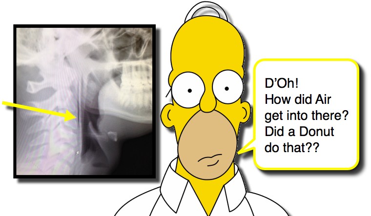
Every so often you encounter a patient that has a finding that catches you by surprise. Like having air in places that should not have air in them. We have discussed spontaneous pneumothorax and traumatic pneumothorax in children as well as how to detect pneumothorax in neonates and how to treat a pneumothorax, but what about pneumomediastinum? As you try to resist the urge to say “D’Oh!” let us consider Pneumomediastinum!
MRI isn’t always easy to obtain in the ED. However, when is it absolutely needed? This post evaluates the indications for emergent MRI.
The Utility of MRI in the Emergency Department Read More »
CT: the donut of truth. Most physicians breathe a little easier sending a patient home with a negative CT abdomen/pelvis. However, the power of x-ray vision doesn’t allow us to turn off our brains. Certain pathologies may have only subtle findings on CT, and others may lend themselves better to other imaging modalities, such as ultrasound. By being aware of these pathologies and how to identify them, we can better recognize patients at risk of a missed diagnosis.
Commonly Missed Findings on CT Abdomen/Pelvis Read More »
CC: Headache
First visit
HPI: 29 year old female with a prior history of headaches, presented with two days of gradual onset, atraumatic, right sided headache that is throbbing in nature. The patient reported heaviness about the eye but no visual changes or disturbances. No neck pain, fevers, chills. She described feeling slightly light-headed but no balance loss. She had a mechanical trip and fall yesterday without head trauma, and her headache had been present for a day prior to the fall.
ROS: otherwise normal.
PMH/PSH: headaches, depression, anxiety, asthma
SH: no smoking, no etoh, no drugs
Allergies: Penicillin (rash)
Pertinent Exam
Vitals: 98.6F, BP: 156/85 P: 101, RR: 16, O2: 98%RA
Gen: A&Ox3, well-developed, well-nourished
HEENT: normocephalic, atraumatic, conjunctiva wnl, EOM wnl, PERRL, normal fundoscopic exam, crisp optic discs, normal ROM neck/supple
Chest: wnl
Abd: wnl
Musculoskeletal: wnl
Neuro: CN2-12 intact, normal reflexes, normal muscle tone, normal coordination
Labs: Serum HCG negative
Imaging: None ordered
ED Course: The patient was believed to be experiencing a migraine headache. She had no evidence of head trauma, no signs of infectious etiology, and had no clinical findings or hx for SAH. She was administered Toradol, IVF and Reglan, and discharged with instructions to follow up with neurology and possibly have an outpatient MRI.
Discharge Dx: Headache […]
Bounceback: An Unrelenting Headache Read More »