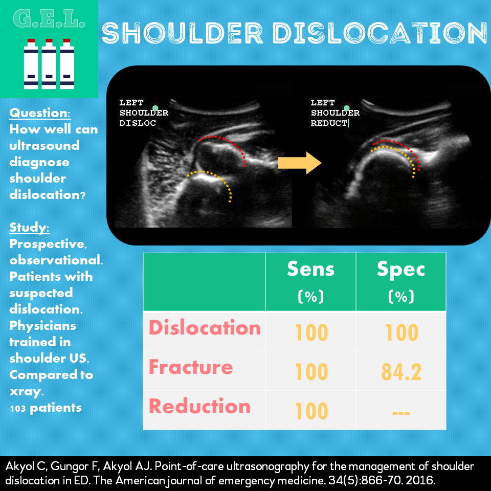
Ultrasound G.E.L. – Ultrasound for Shoulder Dislocation
How does it fare compared to our old friend the xray?
Ultrasound G.E.L. – Ultrasound for Shoulder Dislocation Read More »
How does it fare compared to our old friend the xray?
Ultrasound G.E.L. – Ultrasound for Shoulder Dislocation Read More »

Sepsis is a disease that is prevalent and has a high mortality. We now have a lot of emphasis being placed on early diagnosis of sepsis, early antibiotics, and early source control. We know that ultrasound can find a lot of potential causes for sepsis, and it can potentially save some time in doing so. This article takes a look to see how the diagnosis made with ultrasound compares to a traditional work up – both in time and accuracy.
Ultrasound G.E.L. – POCUS in Sepsis Read More »
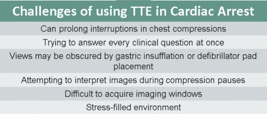
How can you minimize delays in compressions and provide high-quality CPR in cardiac arrest, while using ultrasound? This US Probe post provides you key pearls.
US Probe: Preventing Harmful Delays with POCUS During Cardiac Arrest Read More »
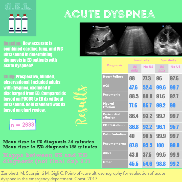
How accurate is combined heart, lung, and IVC ultrasound in determining the diagnosis in an undifferentiated dyspneic patient in the emergency department?
Ultrasound G.E.L. – Acute Dyspnea Read More »
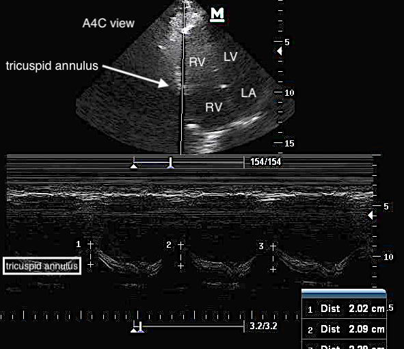
An easy-to-acquire and reproducible measure of right ventricular dysfunction that correlates with outcomes in patients with pulmonary embolism… what’s not to like?
Can you accurately diagnose acute chest syndrome in children with ultrasound? Learn more about this with the latest post from US Probes.

Pericardial tamponade occurs when fluid within the pericardial sac impairs filling of the right-sided chambers, leading to a decrease in cardiac output and hemodynamic compromise. It is neither a clinical nor an echocardiographic diagnosis alone. Rather, the echocardiogram carries diagnostic value and should be performed when there is an elevated pre-test probability for tamponade based on the history and physical exam. Here, we will illustrate the core echocardiographic findings of pericardial tamponade.
US Probe: When Does an Effusion Become Pericardial Tamponade? Read More »
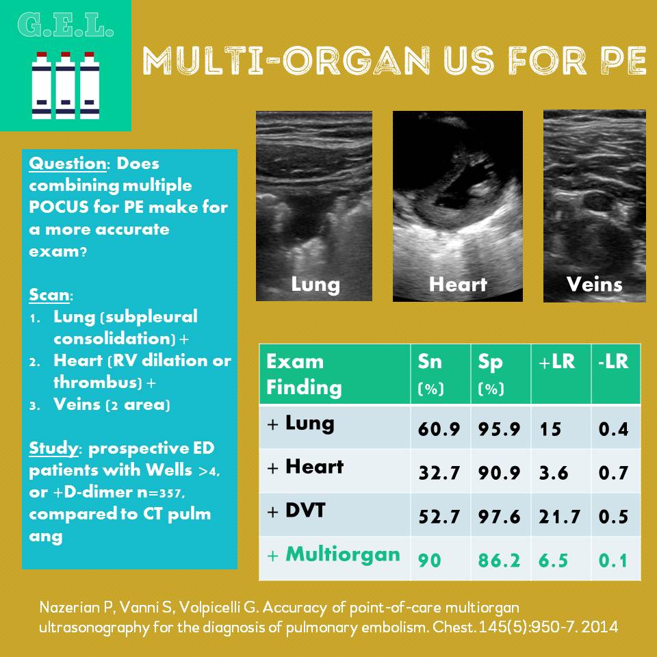
The emDOCs team is happy to be reposting Ultrasound G.E.L. content in their never ending coverage of evidence based ultrasonography (EBU). In this post, does combining multiple POCUS for PE make for a more accurate exam?
Ultrasound G.E.L. – Multiorgan Ultrasound for Pulmonary Embolism Read More »
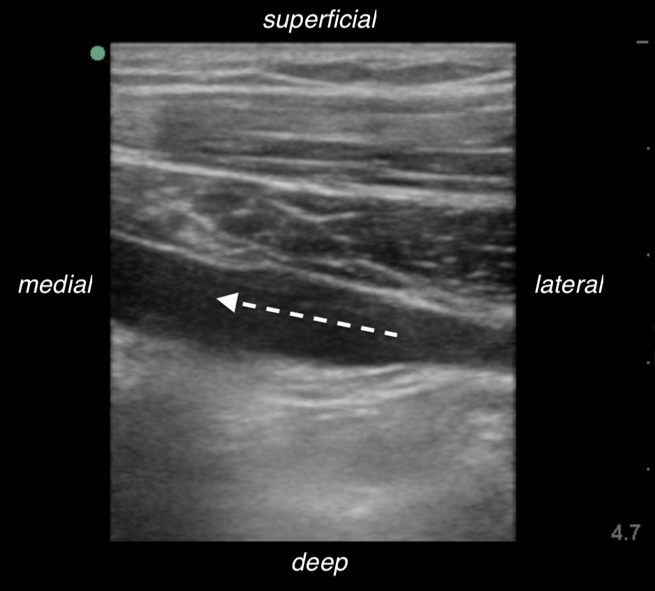
Subclavian/axillary vein catheterization — a not commonly performed ED procedure — using ultrasound guidance is simple to do!
US Probe: Ultrasound Guided Subclavian/Axillary Vein Catheterization Read More »
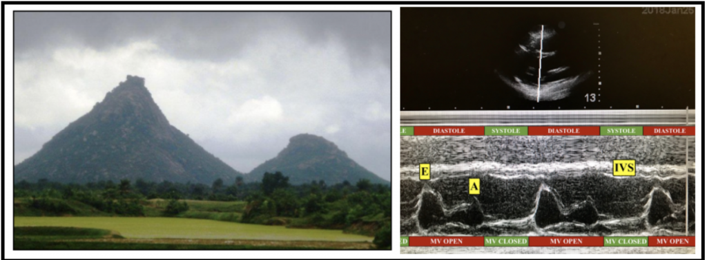
EPSS is a simple, easy to learn tool that allows a quick estimation of left ventricular function. The value of EPSS lies in its objective findings that do not require specialized training for interpretation and utilization, as shown by numerous studies. In patients without mitral or aortic valvular pathology, EPSS can be obtained from a single echocardiography view, providing quantifiable information on heart function within minutes. EPSS > 7mm is typically cited as the cut-off for abnormal ejection fraction (<50%). It offers a further tool for inexperienced emergency physicians that can be used to complement the overall assessment and risk stratification of patients with congestive heart failure.
US Probe: E-Point Septal Separation (EPSS) in the CHF Patient Read More »