General Info / Main Questions
Can ultrasound be used to accurately evaluate shoulder dislocation? Is intra-articular injection as effective as procedural sedation for shoulder reduction?
Ultrasound seems to be finding more and more applications in the emergency setting as training programs, fellowship programs, and even medical schools are integrating ultrasound education into their curriculums. Ultrasound is a non-invasive diagnostic and procedural tool that has become the gold standard and standard of care in some diagnoses and procedures, such as identifying an IUP or placing a central line. It also allows bedside diagnosis in emergent settings and has been studied as a diagnostic tool for shoulder dislocations. Current diagnosis usually consists of the clinical exam and X-ray imaging (Rosen 53: 631). Ultrasound is showing promise as a sensitive diagnostic tool, with 100% sensitivity in one study.2
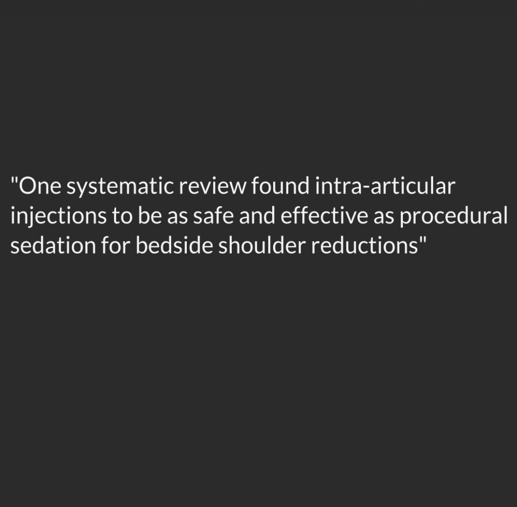
Ultrasound can also be used to for procedural guidance of intra-articular joint injections. One systematic review found intra-articular injections to be as safe and effective as procedural sedation for bedside shoulder dislocation reductions1, and is described in Roberts and Hedges Textbook of Clinical Procedures.
Diagnostic ultrasound
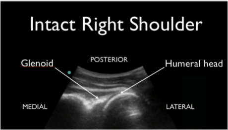
- The ultrasound probe (linear or curvilinear) is positioned posterior to the patient and horizontal to the spine of the scapula.
- Slide the probe laterally to visualize the glenoid and humeral head.
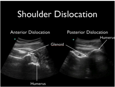
- Normal = humeral head located in the glenoid fossa
- Abnormal = humeral head in the near field (posterior dislocation) or far field (anterior dislocation)
Ultrasound-guided intra-articular injection
- Place ultrasound gel and Tegaderm over the probe
- Sterilize the skin over the shoulder and place local anesthetic with a small bore needle
- Find the glenoid and humeral head as described above
- Using a long axis or in-plane technique guide a 20-gauge spinal needle into the joint and inject 20 mL of 1% lidocaine into the joint space / hemarthrosis
- Wait 10 minutes for anesthetic to take effect
Recap Basics
- Ultrasound can be a fast and safe method to diagnose shoulder dislocation
- Obtain image posteriorly and identify the glenoid and humeral head
- Ultrasound can be used to guide intra-articular analgesia for shoulder reduction
- Long axis guidance into the hemarthrosis with 20-gauge spinal needle
Images
Further Reading / References
- Hunter, B, Wilbur, L MD. Can Intra-articular Lidocaine Supplant the Need for Procedural Sedation for Reduction of Acute Anterior Shoulder Dislocation? Ann Emerg Med 59(6): 513-4; 2012.
- Abbasi, S, et. al. Diagnostic Accuracy of Ultrasonographic Examination in the Management of Shoulder Dislocation in the Emergency Department. Ann Emerg Med 62(2): 170-5; 2013.
- Chilstrom, M. (2013, November 3). Lecture presented at the Essentials of Emergency Medicine Conference, San Francisco, CA.
- http://www.ncbi.nlm.nih.gov/pubmed/23490112
- http://www.ncbi.nlm.nih.gov/pubmed/22944540
- http://www.ncbi.nlm.nih.gov/pubmed/19912129
- http://www.ncbi.nlm.nih.gov/pubmed/19041566
- http://www.ncbi.nlm.nih.gov/pubmed/11904265


1 thought on “Shoulder Ultrasound: Intra-Articular Injection and Reduction”
Pingback: Luxation antérieure de l’épaule | thoracotomie