Authors: Shannon N. Thompson, MD, MAEd (EM Resident Physician, San Antonio, TX) and Erica Simon, DO, MPH, MHA (EM Attending Physician, San Antonio, TX) // Reviewed by: Edward Lew, MD (@elewmd); Alex Koyfman, MD (@EMHighAK); and Brit Long, MD (@long_brit)
Case 1
A 75-year-old female with a history of diabetes mellitus and irritable bowel syndrome presents with one day of abdominal pain with numerous loose, bloody bowel movements. Since the onset of her abdominal pain, the patient has noted a persistent urge to defecate.
VS: HR 82 bpm, RR 14, BP 142/85, SpO2 98% RA, T 98.6°F
Abdominal exam is significant for left lower quadrant tenderness without guarding or rebound.
Case 2
An 80-year-old male with a history of coronary artery disease, diabetes mellitus, and stage IV chronic kidney disease presents with acute onset, severe right lower quadrant abdominal pain. The patient reports nausea but denies fevers, chills, hematochezia, melena, sick contacts, and recent travel.
VS: HR 120, BP 85/63, RR 22, SpO2 99% RA, T 98.7°F
Abdominal exam is significant for tenderness in right lower quadrant and distension.
What is your differential diagnosis for each patient? What are the next steps in your ED evaluation?
Background
Ischemic colitis (IC) is the underlying etiology in 16-24% of acute lower gastrointestinal bleeds (LGIBs).1 As individuals with IC often present with non-specific symptoms, approximately 80% of cases are missed during the initial patient encounter.2 In the ED, IC is correctly diagnosed in only 9% of the cases (when compared to final inpatient diagnoses).2 With a 10-15% mortality rate2 and increasing incidence in the U.S.,3 IC is a diagnosis that should not be missed.
Risk Factors & Pathophysiology
Patients with IC are most often at least 50 years old; female; and have a history of hypertension, vascular disease, chronic kidney disease, and/or diabetes.4,5 IC may also be seen in young persons with collagen vascular diseases, polyangiitis granulomatosis, and amyloidosis.6
While the exact mechanism of IC is often unknown, it is thought to develop from low blood flow to the colon. When compared to the small intestine, the colon has a lower volume of blood supply per surface area. It also relies on collateral circulation in the “watershed” areas (splenic flexure and a portion of the sigmoid colon).7 These areas can be under-perfused in episodes of transient hypotension, and can lead to transmural infarction within 8-16 hours if the ischemia is severe.8 IC is a segmental disease with the splenic flexure the most often affected.5 Additionally, aortic procedures that sacrifice the inferior mesenteric artery also increase the risk of IC, as perfusion of the colon then becomes reliant on collateral flow.9,10
There is some evidence that preceding constipation, irritable bowel syndrome, and the use of constipation-inducing, immunomodulating, or illicit drugs increases the risk of IC.9,11–13 Constipation and constipation-inducing drugs are thought to predispose to IC as distension of the bowel decreases blood supply to the mucosa and increases the risk of ischemia.7 Illicit drugs, particularly amphetamines and cocaine, can cause vasoconstriction, direct endothelial injury, and coagulopathy resulting in low flow states or vascular occlusion.14 Immunomodulators, including interleukin-2, interferon, sodium aurothiomalate, and solumedrol, may increase thrombogenesis leading to an occlusive event that precedes IC.14
IC should be differentiated from mesenteric ischemia in which a disruption of the mesenteric circulation results in ischemia of the small bowel.15 While both can occur in the setting of poor perfusion, acute mesenteric ischemia results in ischemia of the small bowel and acute mesenteric ischemia usually occurs due to occlusion of the superior mesenteric artery.15 Whereas, ischemic colitis affects the colon in the distribution of the inferior mesenteric artery.2,16 Patients with CI are typically older, more likely to present with GI bleeding, and less likely to report abdominal pain as their chief complaint when compared to those with mesenteric ischemia.2 Comparatively, mesenteric ischemia carries a much higher mortality rate of 60-80%.15
Patient Presentation & Differential Diagnosis
Symptoms usually include acute abdominal pain, mild rectal bleeding, and diarrhea.4,9 In cases of isolated right colon ischemia, patients often present with abdominal pain in the absence of hematochezia or melena.5 Abdominal pain was one of the clinical features in approximately 87% of patients with IC. While rectal bleeding was present in 84% and diarrhea in 57% according to a study that retrospectively analyzed hospital stays of patients who were discharged with International Classification of Diseases codes that were consistent with IC.4 Both abdominal pain and rectal bleeding occurred in 72% of the hospitalizations analyzed.4 The differential diagnosis for abdominal pain and bloody diarrhea is broad with infection, diverticulosis, hemorrhoids, neoplasia, inflammatory bowel disease, and vascular ectasia all representing causes of LGIB.3 Distinguishing between these conditions based upon history, physical exam, laboratory testing, and imaging is frequently necessary.
Evaluation
While there is no current American College of Emergency Physicians guideline pertaining to ischemic colitis, an excellent guideline based upon nearly 200 resources has been offered by the American College of Gastroenterology (ACG).9 In this publication, the authors recommend diagnosis based upon the presence of symptoms consistent with IC (e.g. sudden cramping, mild abdominal pain, urgent desire to defecate, and passage of bright red or maroon blood or bloody diarrhea within 24 hours of the onset of symptoms).9 The certainty of this diagnosis is strengthened in the setting of the risk factors discussed above.
Laboratory Studies
The guideline9 also recommends extensive laboratory testing for the determination of prognosis and severity:
- Albumin, amylase, complete blood count, comprehensive electrolyte panel, creatinine kinase, lactate, lactate dehydrogenase (LDH)
- Clostridium difficile toxin assay, stool culture, and stool ova and parasite.
Low hemoglobin, less than 12 g/dl, or albumin levels less than 2.8 g/l, and the presence of acidosis, as indicated by lactate and LDH, indicate a more severe course.9 (Table 1) Stool studies serve to rule out infectious etiologies of the hematochezia which, if present, would decrease the clinical suspicion for IC.
Imaging:
Radiographs: Plain films may be considered for patients presenting with guarding and rebound on abdominal exam or if the patient is unstable for computed tomography. Early in the disease course, air fluid levels suggesting ileus may be visualized. As ischemia and infarction progress, mural thickening from submucosal hemorrhage may appear as “thumbprinting.” Later signs may include pneumatosis coli and perforation (free air under the diaphragm).7
Ultrasound (US): A paucity of literature exists regarding the use of US for the detection of IC. While bowel wall thickening and lack of arterial flow on color doppler may be suggestive, differentiating between the types of colitis and/or malignancy may be difficult.17,18
Computed Tomography (CT): The ACG, recommends a CT of the abdomen with intravenous and oral contrast as the first choice for the diagnosis of IC given its ability to assess the phase (acute, subacute, or chronic) and distribution of the colitis. Alternatively, a multiphasic CTA could be utilized in severe disease (Table 1).9 It also states that a CTA is not necessary if mesenteric ischemia is not a likely diagnosis, as blood flow to the colon has likely normalized by the time of presentation for patients with ischemic colitis. 9 However, given the likely uncertainty in the diagnosis in the undifferentiated ED patient and the difficulty in distinguishing IC from mesenteric ischemia, authors recommend considering CTA first in the ED patient in whom there is concern for IC, provided resources allow. This is supported by a recent article in the British Journal of Radiology which cites the sensitivity of CTA in determining etiology of acute GI bleeding to be above 90% and supports consideration of CTA as a first-line modality.19
Findings of bowel wall thickening, edema, and thumbprinting on CT suggest IC. Colonic pneumatosis and portomesenteric venous gas are more specific and indicate more severe disease.9 Left-sided colonic involvement is most common,5 while right-sided disease is likely to be more severe.20 If the IC is isolated to the right colon on CT, a multiphasic CTA is recommended due to the possibility of the IC resulting from acute mesenteric ischemia with possible involvement of the SMA.9 This can be done prior to discharge once the patient is admitted and does not need to be performed in the ED.9


More CT imaging and descriptions can be found at Radiopaedia.org by this link. Sensitivities and specificities for each modality are not available, likely secondary to the difficulty in confirming the diagnosis of IC.
Isolated right colon ischemia (IRCI) is associated with much poorer outcomes and significant mortality. Patients with IRCI are five times more likely to need surgical intervention and have a twofold increased mortality.20
In the hemodynamically stable patient with IC risk factors and no indications for immediate surgical intervention (hematochezia/melena, peritoneal signs, or evidence of severe disease on imaging) colonoscopy is recommended within 48 hours of presentation to confirm the diagnosis. In this patient population, it is possible that colonic blood flow has returned to baseline prior to imaging, making vascular studies less useful.9
Prognosis & Disposition
Most episodes of IC will resolve spontaneously without specific therapy. However, several factors have been associated with more severe disease and a poorer outcome (Table 1). Factors indicating severe disease include: male gender (odds ratio 3.94), hypotension (systolic blood pressure <90 mmHg, odds ratio 4.45), tachycardia (heart rate >100 beats/min, odds ratio 4.40), abdominal pain without rectal bleeding (more common in isolated right colon ischemia, odds ratio 3.90), BUN >20 mg/dl, Hgb <12 g/dl (odds ratio 4.50), LDH >350 U/I, serum sodium <136 mEq/l (mmol/l) (odds ratio 4.98), WBC >15 cells/cmm (x109/L) with the higher odds ratio associated with increased requirement for surgery and/or mortality.9 (Table 1)

Patients with IC should be admitted for optimization of underlying medical conditions as well as a colonoscopy to confirm the diagnosis.
Management
The management of IC depends on disease severity.
A patient is considered to have severe disease if he or she has more than three of the criteria in Table 1; peritoneal signs on physical exam; pneumatosis coli, portal venous gas, pan-colonic involvement, or IRCI upon imaging.9 If severe disease is suspected prior to imaging, a CTA should be performed instead of CT. Surgery should be emergently consulted for any patient with peritoneal signs, massive bleeding, universal fulminant colitis, or deteriorating clinical condition.9The patient should be started on broad spectrum antibiotics. The recommendation is for anaerobe and gram-negative coverage with metronidazole or clindamycin combined with a fluoroquinolone, aminoglycoside, or 3rd generation cephalosporin.21 Antimicrobial therapy is believed to decrease bacterial translocation in the injured bowel in the setting of ischemia and reperfusion injury; although no definitive evidence exists for its benefit, nor has a specific regimen been shown to be most eficacious.9 Depending upon the surgical consultation, disposition will be immediately to the operating room or to the intensive-care unit (ICU).9
Patients with moderate disease have typical symptoms of IC and 1-3 of the factors in Table 1 indicating more severe disease. They also have segmental colitis without involvement of the right colon on CT imaging. For these patients, antibiotics should be considered. Underlying cardiovascular abnormalities and fluid deficits should be corrected, and the patient should be admitted for supportive care, correction of underlying etiologies, and colonoscopy.10
Those with typical symptoms of IC with CT imaging similar to that of moderate disease without risk factors in Table 1 are considered to have mild disease. The treatment for this is admission for observation, diagnostic colonoscopy, and supportive care.
In resource-limited or austere environments, care should be taken to identify moderate and severe disease. These categories would likely benefit from transfer or evacuation for CT and/or colonoscopy. Mild disease is likely to be self-limiting, and therefore may be considered for admission for observation with symptomatic treatment for the presumptive diagnosis.
Case 1 Conclusion:
In the first case, the patient’s labs return without any of the abnormalities in Table 1, and the CT does not show pan-colonic involvement or IRCI. The patient is considered to have mild disease and is admitted for observation and colonoscopy.
Case 2 Conclusion:
The patient in Case 2 requires immediate resuscitation. This patient has severe disease and likely IRCI, given the abdominal pain without rectal bleeding and CKD, which have both been associated with IRCI.5 An emergent surgical consultation was obtained and the patient is admitted to the ICU. A CTA is later performed to rule out involvement of the superior mesenteric artery and mesenteric ischemia.
Pearls
- Consider ischemic colitis as the possible cause of acute-onset abdominal pain with rectal bleeding in patients age 50 and older with cardiovascular morbidities.
- For patients with peritoneal signs, radiographs should be considered for evaluation of ileus (air fluid levels), thumbprinting, pneumatosis coli, or pneumoperitoneum.
- Consider ordering a CTA as first-line imaging, especially if there is any suggestion of mesenteric ischemia, right sided tenderness without hematochezia, or if you suspect severe disease. If resources do not allow, order a CT with oral and IV contrast. If IRCI is incidentally found on initial CT, then CTA can be deferred to the inpatient setting and obtained prior to discharge.
- Give antibiotics in suspected ischemic colitis if the patient has any of the factors associated with severe disease.
- Place an emergent surgical consultation for any patient with peritoneal signs on physical exam or pneumatosis coli, portal venous gas, pan-colonic distribution, or isolated right-colon ischemia on imaging.
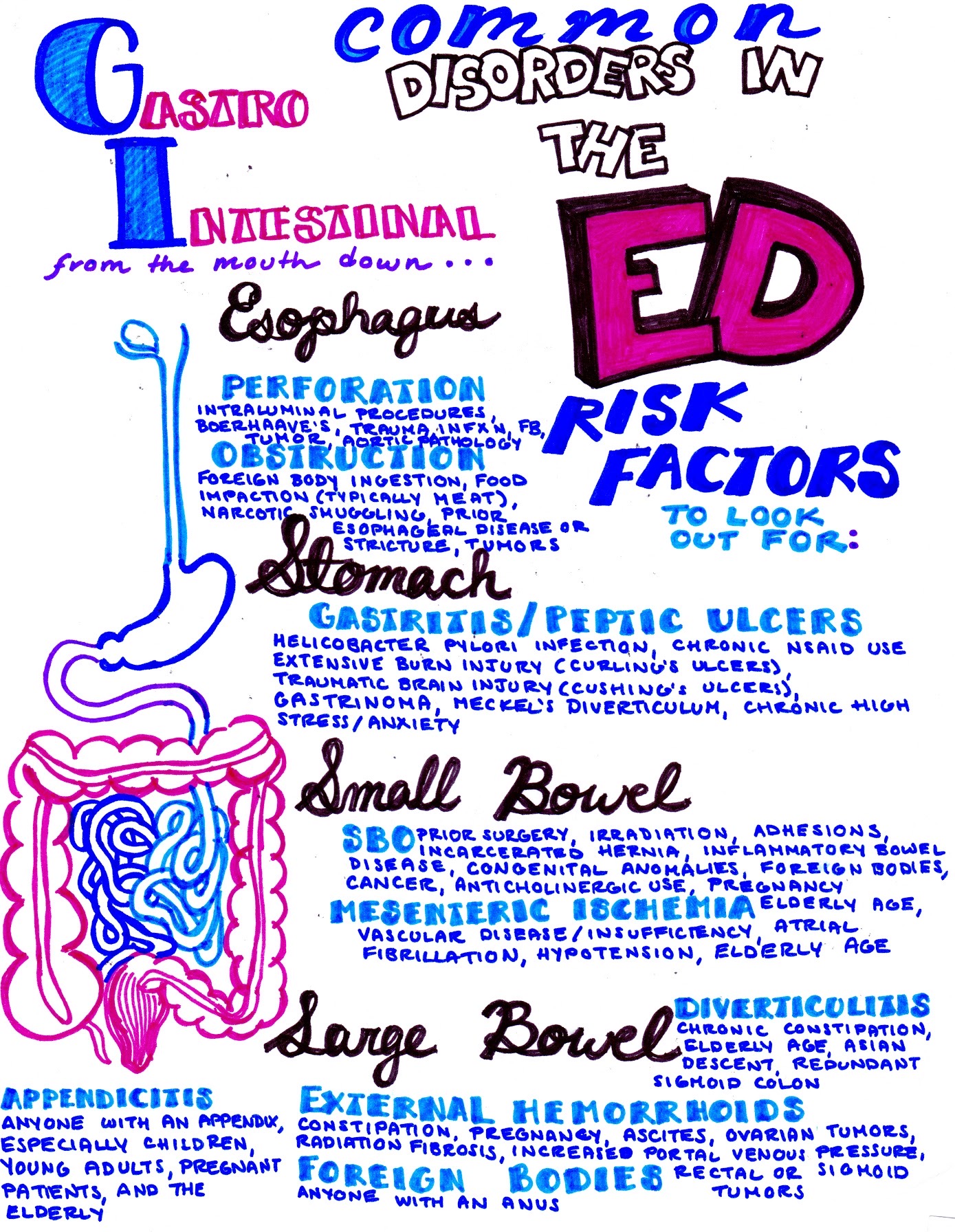
From Dr. Katy Hanson at Hanson’s Anatomy:


References/Further Reading
- Hreinsson JP, Gumundsson S, Kalaitzakis E, Björnsson ES. Lower gastrointestinal bleeding: incidence, etiology, and outcomes in a population-based setting. Eur J Gastroenterol Hepatol. 2013;25(1):37-43. doi:10.1097/MEG.0b013e32835948e3
- Ullery BS, Boyko AT, Banet GA, Lewis LM. Colonic ischemia: an under-recognized cause of lower gastrointestinal bleeding. J Emerg Med. 2004;27(1):1-5. doi:10.1016/j.jemermed.2003.11.022
- Gayer C, Chino A, Lucas C, et al. Acute lower gastrointestinal bleeding in 1,112 patients admitted to an urban emergency medical center. Surgery. 2009;146(4):600-607. doi:10.1016/j.surg.2009.06.055
- Longstreth GF, Yao JF. Epidemiology, clinical features, high-risk factors, and outcome of acute large bowel ischemia. Clin Gastroenterol Hepatol Off Clin Pract J Am Gastroenterol Assoc. 2009;7(10):1075-1080.e1-2; quiz 1023. doi:10.1016/j.cgh.2009.05.026
- Brandt LJ, Feuerstadt P, Blaszka MC. Anatomic patterns, patient characteristics, and clinical outcomes in ischemic colitis: a study of 313 cases supported by histology. Am J Gastroenterol. 2010;105(10):2245-2252; quiz 2253. doi:10.1038/ajg.2010.217
- Goldblum J. “Large Bowel” Rosai and Ackerman’s Surgical Pathology. 11th ed. New York, NY: Elsevier; 2018.
- FitzGerald JF, Hernandez III LO. Ischemic Colitis. Clin Colon Rectal Surg. 2015;28(2):93-98. doi:10.1055/s-0035-1549099
- Haglund U, Bulkley GB, Granger DN. On the pathophysiology of intestinal ischemic injury. Clinical review. Acta Chir Scand. 1987;153(5-6):321-324.
- Brandt LJ, Feuerstadt P, Longstreth GF, Boley SJ. ACG Clinical Guideline: Epidemiology, Risk Factors, Patterns of Presentation, Diagnosis, and Management of Colon Ischemia (CI). Am J Gastroenterol. 2015;110(1):18. doi:10.1038/ajg.2014.395
- Van Damme H, Creemers E, Limet R. Ischaemic colitis following aortoiliac surgery. Acta Chir Belg. 2000;100(1):21-27.
- Walker AM, Bohn RL, Cali C, Cook SF, Ajene AN, Sands BE. Risk factors for colon ischemia. Am J Gastroenterol. 2004;99(7):1333-1337. doi:10.1111/j.1572-0241.2004.21436.x
- Park CJ, Jang MK, Shin WG, et al. Can we predict the development of ischemic colitis among patients with lower abdominal pain? Dis Colon Rectum. 2007;50(2):232-238. doi:10.1007/s10350-006-0753-5
- Cole JA, Cook SF, Sands BE, Ajene AN, Miller DP, Walker AM. Occurrence of colon ischemia in relation to irritable bowel syndrome. Am J Gastroenterol. 2004;99(3):486-491. doi:10.1111/j.1572-0241.2004.04097.x
- Hass DJ, Kozuch P, Brandt LJ. Pharmacologically mediated colon ischemia. Am J Gastroenterol. 2007;102(8):1765-1780. doi:10.1111/j.1572-0241.2007.01260.x
- Clair DG, Beach JM. Mesenteric Ischemia. Campion EW, ed. N Engl J Med. 2016;374(10):959-968. doi:10.1056/NEJMra1503884
- Washington C, Carmichael JC. Management of Ischemic Colitis. Clin Colon Rectal Surg. 2012;25(4):228-235. doi:10.1055/s-0032-1329534
- Ripollés T, Simó L, Martínez-Pérez MJ, Pastor MR, Igual A, López A. Sonographic findings in ischemic colitis in 58 patients. AJR Am J Roentgenol. 2005;184(3):777-785. doi:10.2214/ajr.184.3.01840777
- Danse EM, Jamart J, Hoang P, Laterre PF, Kartheuser A, Van Beers BE. Focal bowel wall changes detected with colour Doppler ultrasound: diagnostic value in acute non-diverticular diseases of the colon. Br J Radiol. 2004;77(923):917-921. doi:10.1259/bjr/18038687
- Wortman JR, Landman W, Fulwadhva UP, Viscomi SG, Sodickson AD. CT angiography for acute gastrointestinal bleeding: what the radiologist needs to know. Br J Radiol. 90(1075). doi:10.1259/bjr.20170076
- Sotiriadis J, Brandt LJ, Behin DS, Southern WN. Ischemic Colitis Has a Worse Prognosis When Isolated to the Right Side of the Colon. Am J Gastroenterol. 2007;102(10):2247-2252. doi:10.1111/j.1572-0241.2007.01341.x
- Brandt LJ, Feuerstadt P. Beyond Low Flow: How I Manage Ischemic Colitis. J Gastroenterol. 2016;111(12):1672-1674. doi:10.1038/ajg.2016.456


1 thought on “Ischemic Colitis: ED Presentations, Evaluation, and Management”
Pingback: Quiz 54, December 13th, 2019