
Authors: Joseph R. Peters, DO, RDMS, FACOEP, FACEP, FACOI (EM Attending Physician, OSF St. Francis Medical Center); Carolina Hartje, DO (EM Resident Physician, OSF St. Francis Medical Center) // Reviewed by: Stephen Alerhand, MD; Alex Koyfman, MD (@EMHighAK); Brit Long, MD (@long_brit)
Introduction
There are an estimated 300,000-600,000 Americans who develop venous thromboembolisms (VTE) each year, including deep venous thromboembolisms (DVT) and pulmonary embolism (PE).1-3 VTE is responsible for hospitalizing over 250,000 Americans every year, and there are an estimated 100,000 deaths annually associated with these conditions. 1-3 As many as 25% of acute PE cases present as sudden cardiac death.4 In an emergency department (ED) presentation of cardiac arrest, the diagnosis of PE is challenging without the use of CT angiography. Point-of-Care-Ultrasound (POCUS) is a bedside modality that can assist Emergency Physicians (EPs) in differentiating PE from other causes of cardiac arrest.
Case
A 25-year-old-female presented to the ED in cardiac arrest. Paramedics reported that coworkers activated them after the patient was discovered unconscious with labored breathing. There was no other known medical history, and they found the patient recumbent in her desk chair pulseless, not breathing, with a fixed leftward gaze. EMS verbalized concern to EPs that an “intracranial bleed” may have precipitated the event. Advanced cardiac life support (ACLS) had been initiated and on arrival at the ED, the patient was found to have Pulseless Electrical Activity (PEA). The Accu-Chek was 214, and there was no response to empiric Narcan administration. This history heightened EPs concern for a massive PE causing labored breathing leading to brain anoxia and gaze deviation. Given either scenario was plausible, differentiating the etiology of this presentation would be challenging given that the arrest-state precluded the use of CT scan. Time was of the essence, the patient was intubated, ACLS measures were continued, and POCUS was performed. The POCUS findings enabled EPs to confidently recognize findings consistent with hemodynamically unstable (formerly sub-massive or massive) PE. Consultants were rapidly mobilized, and therapy was administered confidently within minutes of arrival.
Multiorgan POCUS
The diagnostic power of POCUS often resides in combining multiple ultrasound exams. Although POCUS exams are learned individually, scanning condition-related anatomic areas provides EPs with critical information, improving the opportunity to rule-in a diagnosis more confidently. In one study multi-organ POCUS of the heart, lungs, and lower extremities was 90% sensitive and 86% specific for the diagnosis of PE, compared to individual POCUS exams alone.5 In this case, multi-organ POCUS findings consistent with PE enabled EPs to diagnose the cause of cardiac arrest in a scenario where the patient was too unstable to obtain immediate CT.
The Pathophysiology
Blood clots classically form in the deep veins of the proximal legs and then embolize into the pulmonary arteries.7,8,16 Approximately 45% of patients with verified PE are found to have a residual proximal DVT.7 In one study, DVT was found in 70% of patients with proven PE.16 DVTs often include unilateral leg pain, swelling, erythema, and pain in the calf with dorsiflexion of the foot, known as Homan’s sign. However, many DVTs are asymptomatic before embolization, as in our patient.6,7
Once a DVT embolizes into the pulmonary arterial circulation, symptoms develop when more than 20% of their pulmonary vasculature is occluded.7 The classic symptoms of PE include dyspnea, pleuritic chest pain, cough, and less commonly syncope or arrest. The degree of obstruction of blood flow in the pulmonary arteries contributes to which symptoms and presentations occur. Some experts explain that the pulmonary artery pressure does not rise significantly unless greater than 30% of the pulmonary arteries are obstructed.8,16 They explain that POCUS findings of RV strain may not be visualized with POCUS echocardiography until more than 50% of the pulmonary artery outflow track is obstructed.8,16 For this reason, POCUS echo findings may not be present unless a hemodynamically unstable PE is present. This likely explains why some individuals remain entirely asymptomatic with normal POCUS findings when flow obstruction is insignificant. In one study, PE was found in 32% of patients with diagnosed DVT despite not having any PE symptoms.9
POCUS Echocardiography
Echocardiography is an important POCUS exam in cardiac arrest patients. As per the American Society of Echocardiography-American College of Emergency Physicians (ASE-ACEP) Consensus statement, the ability to assess global left ventricular function, to detect pericardial effusions, and to assess for right heart dilatation (chamber sizes) are within the scope of clinicians and can help answer critical patient management questions.10
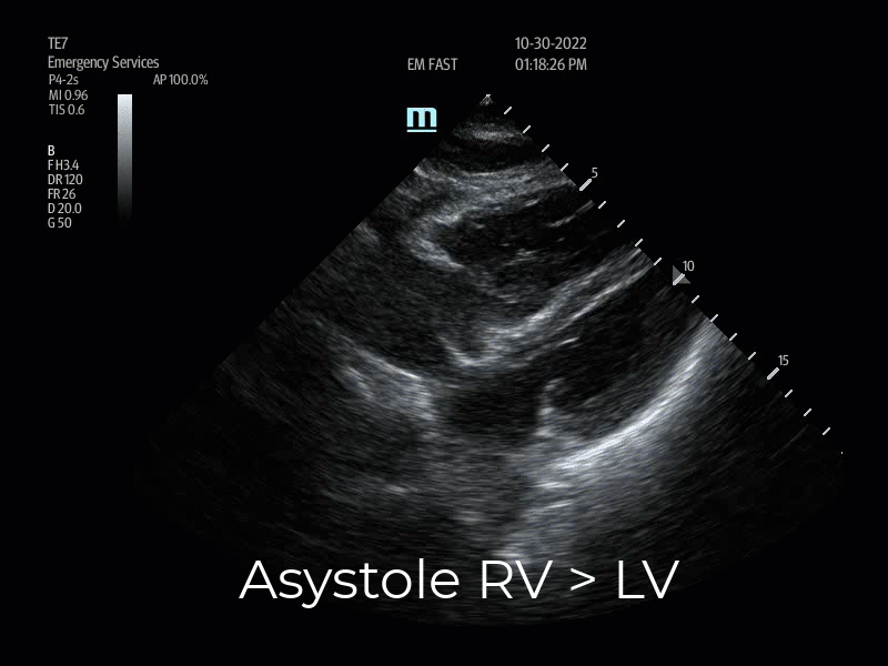
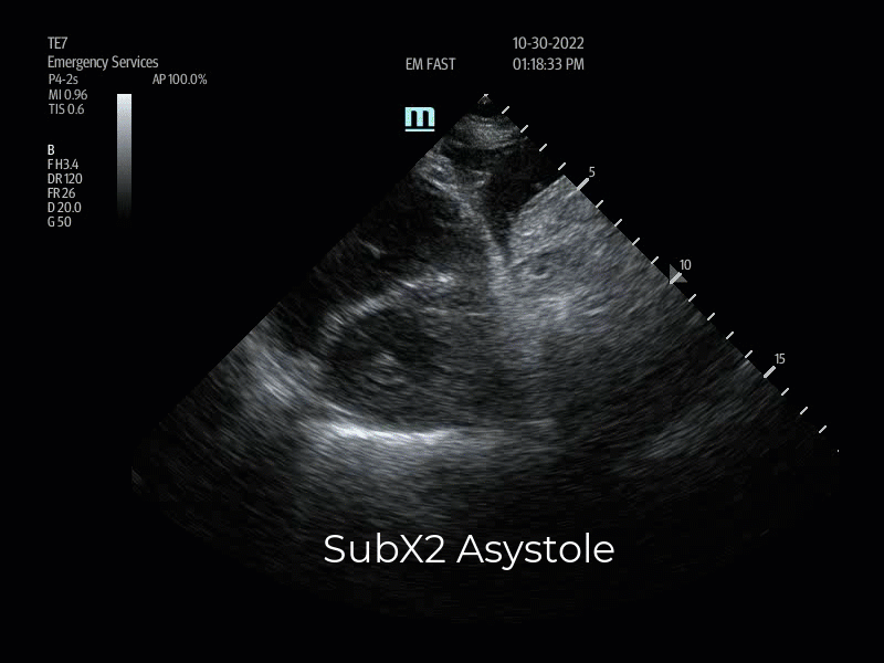
A subxiphoid 4-chamber view (SubX4) was obtained in a 66-year-old cardiac arrest patient who was eventually diagnosed with PE by CT angiography (Vid. 1). At first pulse check, EPs ascertained the patient had no LV function (asystole), no pericardial effusion, but did recognize that the RV cavity appeared subtly larger than the LV cavity, indicating possible RV strain. These findings were also noted at the second pulse check utilizing the subxiphoid 2-chamber (SubX2) view, imaging the RV and LV in short axis from the xiphoid region (Vid 2). RV chamber size alone is not enough information to rule-in a PE as RV cavity enlargement can be visualized in other conditions such as pulmonary hypertension, RV infarct, COPD and cardiac arrest from multiple causes.10,11
Gottlieb et al discusses the reliability of right ventricular dilation for suspected PE in cardiac arrest, highlighting that EPs need to be aware of the following: “Current evidence suggests identifying RV strain in an arrest patient may be less reliable as both animal and human studies have demonstrated that RV dilation is frequently seen in patients with cardiac arrest from an array of causes including arrhythmias, respiratory failure, and circulatory failure in the absence of PE. RV dilation appears to be progressive as cardiac arrest continues and should not be used in isolation to diagnose PE. However, RV dilation may be more suggestive of PE if performed early in the cardiac arrest with a consistent history (sudden chest pain or dyspnea) or in the presence of a DVT. As RV dilation can be seen in chronic pulmonary hypertension, EPs should also evaluate the RV free wall thickness. A thin wall (diameter <5mm) is suggestive of an acute etiology”. 11
With return to spontaneous circulation (ROSC), SubX4 continued to reveal concern for RV strain as the RV cavity appeared equal to slightly more dilated than the underfilled LV (Vid 3). A POCUS DVT exam was performed, and no DVTs were identified in the patient’s lower extremities. However, as shock resolved and hemodynamic stability improved, RV strain and underfilling of the LV remained apparent 30 minutes after ROSC (Vid 4). With this information, in the context of the history of severe sudden onset of dyspnea followed by cardiac arrest, EPs were prompted to obtain CT angiography revealing multiple bilateral PEs, but no saddle embolus.


Normal appearance of the RV & LV chambers
Compared to the LV, the RV is a low-pressure pump with thin walls.9 The normal RV:LV cavity diameter ratio is 0.67-1, and therefore the normal RV cavity appears approximately 1/3rd smaller than the LV cavity in any view of the heart (Vids 5,6,7,8).15 The lateral free wall of the RV appears thin (<5 mm).11 The two ventricles share an interventricular septum which during the cardiac cycle normally remains in a neutral position despite the RV:LV pressure differences (Vids 5,6,7,8).
In the long axis views of the heart, RV function is recognized by the motion of the tricuspid valve annulus moving toward the apex of the heart. This is known as the Tricuspid Annular Plane Systolic Excursion (TAPSE). The RV is different from the LV in that more of its contractile motion is in a longitudinal plane which is a consequence of the RV having more myofibers oriented in an apex-to-base direction.12 EPs with POCUS Echo experience and familiar with normal appearing TAPSE can qualitatively estimate TAPSE visually, especially in cardiac arrest evaluation where time is of essence. The AP4, SubX4, and the Parasternal Long Axis View (PSLAX) views can be used to visually estimate TAPSE. TAPSE motion is RV afterload dependent and correlates closely with RV ejection fraction measured by radionuclide angiography.13 TAPSE is quantitatively measured in the Apical 4 chamber view (AP4) with m-mode to evaluate RV systolic function.12 The lateral annulus of the tricuspid valve is the visual reference at the base of the RV used to measure the longitudinal motion toward the apex (Vid 5, 7, 8).12 Measurements less than 16 mm are considered indicative of poor RV function and correlates with increased mortality.14 Multiple conditions can affect RV function including PE, RV infarct, pulmonary hypertension, cardiomyopathy, and cardiac arrest, so TAPSE is not specific for PE.8,10,11
In the parasternal short axis view (PSSAX), the normal RV cavity appears as a parasitic structure attached to the septal wall of the LV. The RV cavity again appears approximately 1/3rd smaller than the LV. The septal wall of the LV always maintains it’s a rounded shape under normal conditions, referred to the “O-sign” (Vid 6).


Features of RV strain
Right heart strain (RV cavity > LV cavity) alone is not sensitive for the diagnosis of PE as there are several causes. Alerhand et al described ten individual echocardiographic findings of RV strain that suggest PE.15 EPs would benefit to be familiar with all ten findings, however advanced POCUS expertise and doppler ultrasound skill is required to perform six of the ten. In crisis care situations, the quantitative exam finds may be challenging and time consuming to implement. Our case highlights how four of the ten qualitative POCUS findings (RV:LV ratio, qualitatively reduced TAPSE, paradoxical septal wall motion toward the LV, and McConnell’s sign) suggest PE as the etiology of a cardiac arrest patient. These imaging windows require only the basic POCUS skill and can be rapidly implemented in the evaluation of cardiac arrest patients. During this resuscitation two different ultrasound machines were used to capture images due to a malfunction with the first machine after initial images were captured.
Upon arrival, our 25-year-old patient was in PEA arrest. She was not breathing, pulseless, and bradycardic on the monitor. After intubation and ACLS measures, ROSC was obtained briefly. POCUS PSLAX was performed. (Vid 9). The first concern identified was that the RV cavity appeared qualitatively greater in diameter than the LV cavity suggesting RV strain. In hemodynamically unstable PE this finding occurs due to the acute increase in pulmonary vascular resistance (PVR), or RV afterload.8,16 Blood filling the RV is not completely ejected due to pulmonary artery obstruction resulting in increased RV pressure, cavity enlargement, and failure. It was also recognized that the LA and LV appeared underfilled due to lack of preload, as blood could not get through the lung circuit to fill the left chambers. A hyperdynamic LV is defined with POCUS by the lateral wall of the LV nearly touching or touching the septum, indicating low preload.10 When a low preload is present, low cardiac output is present, indicated in this video by the small diameter of the descending aorta. Comparing Vids. 9 and 5 visually highlight the differences. Acute RV failure due to acute pressure overload is the primary cause of death in severe PE.16
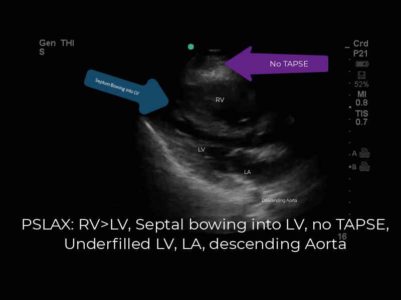
Vid 9. PSLAX: RV Strain, loss of TAPSE, paradoxical septal motion, and underfilled LV, LA, descending aorta.
In the setting of acute PE, complex hemodynamic changes affect right ventricular function.14 Mechanical obstruction of the pulmonary arteries and the release of vasoconstrictive mediators may cause abrupt increases in pulmonary vascular resistance and RV afterload.14 When RV pressure elevates severely, the lateral wall is under extreme tension creating reduced TAPSE motion, recognized as the base of the RV is not moving toward the apex (Vid 9). If RV pressure is extreme, it can exceed LV pressure causing paradoxical septal wall motion toward the LV as well. In this long axis view of this heart, the septum is shifting toward the smaller LV rather than remaining in a neutral position and there is no TAPSE visible (Vid. 9). Bowing of the septum toward the LV can be misinterpreted as normal septal contraction. However, normal septal contraction should only thicken the septum during systole rather than shifting it toward the LV (Vids 5,9). To verify this paradoxical septal motion toward the LV, incorporating the PSSAX view reveals that the normal “O-shape” of the LV is pressure-flattened into a “D-shaped” LV, indicating elevated RV pressure > LV pressure. Flattening of the septal wall creates the “D-sign” in the PSSAX window (compare Vids 6,10).
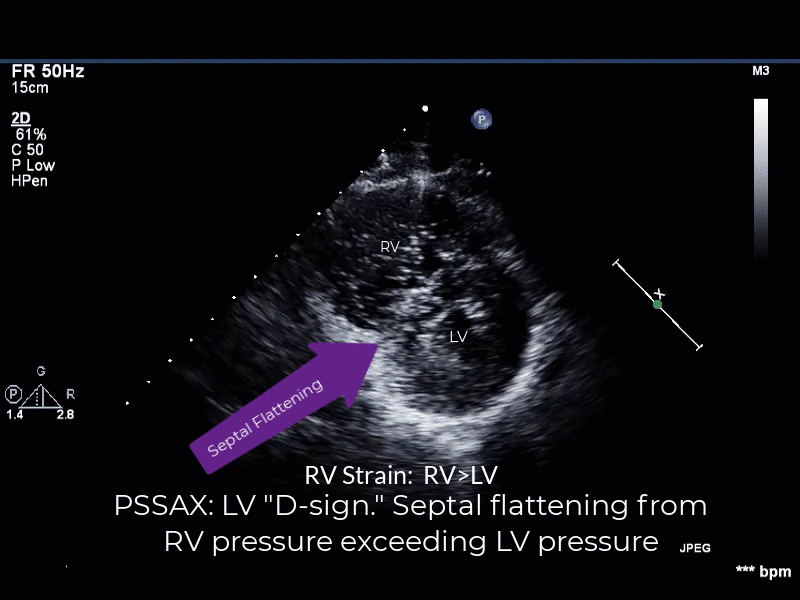
Vid 10. RV Cavity > LV Cavity with septal flattening, “D-sign”. RV pressures > LV pressures.
The AP4 chamber view also indicated that the RV cavity > LV cavity, indicating RV strain (Vid 11). Additional findings supporting obstructive shock include the RA > LA, septal bowing toward the LV, low preload in the LV and LA, and reduced TAPSE motion indicating elevated right sided pressures due to RV outflow obstruction. Recognize also that the normal apical dominance of LV is not present (Compare Vids 11 and 6). Instead, the RV is apically dominant due to the severe pressure overload creating RV enlargement. Severe RV pressure is also noted by the presence of McConnell’s sign, which is visualized as an akinetic right ventricle lateral wall with hyperkinetic or preserved function of the apex.10 All findings consistent with RV failure caused by severe PE.

Vid 11. AP4 RV strain: McConnel’s sign, no TAPSE, RV>LV, septal bowing towards the LV.
Plethoric IVC
The IVC can be considered a clinical barometer of pressures in the RA. When RV pressures elevate, RA pressures increase as well given forward blood flow is obstructed. Identification of a dilated IVC lacking respiratory variation (Plethoric IVC) supports the finding of elevated right heart pressures (Vid 12). There are multiple causes of this finding including positive pressure ventilation, cardiomyopathy, chronic pulmonary hypertension, valvular disease, pericardial tamponade, and PE. POCUS echo often differentiates these etiologies. In our case, the patient was intubated when the IVC view was performed. Positive pressure ventilation was likely a contributor to the finding of plethoric IVC. In pre-intubation patients however, a plethoric IVC in addition to the identification of RV strain, lack of TAPSE, paradoxical septal motion would be confirmatory of pathologically elevated right heart pressures, most likely due to severe PE.
The “IVC inlet view” is an exam that also can visualize Clot-In-Transit (CIT), CIT was not identified in this patient.

Vid 12. IVC inlet view: Plethoric IVC.
Clot-In-Transit (CIT)
Clot-in-transit (CIT) refers to a noticeable, free-floating thrombus inside the right-side chambers of the heart, although a thrombus in the superior vena cava (SVC) or IVC may also fall under this heading.18 About 4% of patients suffering from a PE have a floating embolus present in the right heart, a finding that is associated with a mortality rate of more than 40%.18 EPs also need to be aware that cardiac thrombus formation can be the consequence of forward blood flow ceasing in patients with cardiac arrest.19 Approximately 90% form in <6 min, in accordance with in vivo clotting times (6–10 min).19 Differentiating the etiology of a cardiac thrombus may be challenging. However, if CIT is associated with acute PE, then echocardiography will typically show evidence of right heart strain: interventricular septum bowing into the LV, systolic dysfunction of the RV (loss of TAPSE), and McConnell’s sign.18
Right heart clots can be classified into three categories based on their morphology. Type A is the most common type and occurs when a DVT breaks free and floats up to the right side of the heart.18 It carries the greatest risk of embolization into the pulmonary circuit as it moves freely inside the heart chambers and has an elongated, wormlike form.18 In Type B the atrium or ventricle is assumed to be the source of the thrombus and appears oval, with a broad base, and is adhered to the chamber wall.18 Type C thrombi are uncommon clots that look like cardiac myxomas but may have features of both Type A and B.18,20

Vid 13. Type A CIT. PSLAX RA-RV and AP4 views.
POCUS DVT Exam
Given initial clinical concern that PE was the cause of this cardiac arrest, while ACLS measures were in progress, a POCUS DVT exam was performed. A DVT was visible in the left femoral vein at the Saphenofemoral Junction (SFJ) and distal. It was also noted that the laterally visualized femoral artery was small and lacked blood flow, consistent with the arrest state (Vid 14). This finding combined with POCUS echo findings indicated that severe PE was the cause of the arrest.

Vid 14. DVT Left Saphenofemoral Junction. Under perfused femoral artery.
During a short period of ROSC an ECG was performed. The findings of RV strain on the ECG also supported the POCUS echo findings in this case. Sinus tachycardia with RBBB, ST segment depressions and T-wave inversions in V1-4 (Img. 1).

Img 1. EKG RV strain.
Failed Thrombolysis
Unfortunately, this patient remained clinically unstable after thrombolysis, re-arresting multiple times while in the ED. One study found that 8% of patients with hemodynamically unstable PE do not respond to thrombolysis.17 Patients who were unresponsive to thrombolysis were prospectively defined as patients with both persistent clinical instability and echocardiographic criteria of RV dysfunction.17 The combination of the patient’s history, PEA arrest, and POCUS findings provided the information needed to administer appropriate therapy and to consult the cardiovascular surgeon early who was at bedside within the first hour of her arrival. Empowered by POCUS findings, the patient was taken directly to the OR for rescue surgical embolectomy (Img 2).

Img 2. Emboli from the pulmonary arteries post-thrombolysis.
Summary
This case exemplifies the value of POCUS in cardiac arrest patients in the ED. Without POCUS, EPs were faced with critical decisions based on history and physical exam alone, as the patient was too unstable to leave the ED for CT scan. Multi-organ POCUS findings empowered EPs to provide rapid therapy and mobilize consultants immediately. EPs should be aware that in cardiac arrest, the POCUS findings of RV strain, poor TAPSE motion, paradoxical septal motion toward the LV, plethoric IVC, and lower extremity DVT may be identifiable in patients who present with severe PE. POCUS is a bedside modality indispensable for making critical decisions when advanced testing is not an option due to patient instability.
References/Further Reading
- Jaff MR, McMurtry S, Archer SL, Cushman M, Goldenberg N, Goldhaber SZ. Management of Massive and Submassive Pulmonary Embolisms, Iliofemoral Deep Vein Thrombosis, and Chronic Thromboembolic Pulmonary Hypertension. Circulation. 2011 Mar;123(16):1788-1830. doi: 10.1161/CIR.0b013e318214914f. PMID: 21422387.
- Office of the Surgeon General (US); National Heart, Lung, and Blood Institute (US). The Surgeon General’s Call to Action to Prevent Deep Vein Thrombosis and Pulmonary Embolism. Rockville (MD): Office of the Surgeon General (US); 2008. SECTION I: Deep Vein Thrombosis and Pulmonary Embolism as Major Public Health Problems. https://www.ncbi.nlm.nih.gov/books/NBK44181/
- Beckman MG, Hooper WC, Critchley SE, Ortel TL. Venous thromboembolism: a public health concern. Am J Prev Med. 2010 Apr;38(4 Suppl):S495-501. doi: 10.1016/j.amepre.2009.12.017. PMID: 20331949.
- Heit JA. The epidemiology of venous thromboembolism in the community. Arterioscler Thromb Vasc Biol. 2008 Mar;28(3):370-2. doi: 10.1161/ATVBAHA.108.162545. PMID: 18296591; PMCID: PMC2873781.
- Nazerian P, Vanni S, Volpicelli G, Gigli C, Zanobetti M, Bartolucci M, Ciavattone A, Lamorte A, Veltri A, Fabbri A, Grifoni S. Accuracy of point-of-care multiorgan ultrasonography for the diagnosis of pulmonary embolism. Chest. 2014 May;145(5):950-957. doi: 10.1378/chest.13-1087. PMID: 24092475.
- Marino, P. L. Marino’s the ICU book, Wolters Kluwer Health, pgs 105-109, 4th ed., 2014.
- Tintinalli, J.E. Tintinalli’s Emergency Medicine: A Comprehensive Guide, McGraw-Hill, pgs 389-396, 9th ed., 2020.
- Ferre R., Boyd J., VUMC Emergency Medicine, “RV Strain & TAPSE” YouTube Video https://youtu.be/6_frVD9Y1pk?si=GdUw7oCFIPe_rfoV
- Farcy et al. Critical Care Emergency Medicine, McGraw-Hill, pgs 129-132, 2nd ed., 2017.
- Dawson, Mallin, Hwang J., Introduction to Bedside Ultrasound, iBook Ch 2, Basic Cardiac, Pg 27.
- Gottlieb M, Sundaram T, Olszynski P, Atkinson P. Just the facts: point-of-care ultrasound in cardiac arrest. CJEM. 2022 Sep;24(6):579-581. doi: 10.1007/s43678-022-00336-7. Epub 2022 Jun 30. PMID: 35771485.
- Ho SY, Nihoyannopoulos P. Anatomy, echocardiography, and normal right ventricular dimensions. Heart. 2006;92 Suppl 1(Suppl 1):i2-i13
- Feger J, Weerakkody Y, Tricuspid annular plane systolic excursion. Reference article, Radiopaedia.org (Accessed on 07 Feb 2024) https://doi.org/10.53347/rID-86469
- Lobo JL, Holley A, Tapson V. Prognostic significance of tricuspid annular displacement in normotensive patients with acute symptomatic pulmonary embolism. J Thromb Haemost. 2014;12(7):1020-7.
- Alerhand S, Sundaram T, Gottlieb M. What are the echocardiographic findings of acute right ventricular strain that suggest pulmonary embolism? Anaesth Crit Care Pain Med. 2021 Apr;40(2):100852. doi: 10.1016/j.accpm.2021.100852. Epub 2021 Mar 26. PMID: 33781986.
- Vyas V, Goyal A. Acute Pulmonary Embolism. [Updated 2022 Aug 8]. In: StatPearls [Internet]. Treasure Island (FL): StatPearls Publishing; 2024 Jan-. Available from: https://www.ncbi.nlm.nih.gov/books/NBK560551/
- Meneveau, Nicolas, et al. “Management of Unsuccessful Thrombolysis in Acute Massive Pulmonary Embolism.” Chest, vol. 129, no. 4, 2006, pp. 1043–50, https://doi.org/10.1378/chest.129.4.1043
- Patel AN, Amrutiya RJ, Manvar BN. A Proposed Approach for the Management of Clot-in-Transit. Cureus. 2022 Aug 27;14(8):e28481. doi: 10.7759/cureus.28481. PMID: 36176887; PMCID: PMC9512516.
- de Gregorio C, Stanzione A. Cardiac Thrombus Formation During Cardiopulmonary Resuscitation for Cardiac Arrest: Is It Time for Ultrasound-Enhanced Algorithms? J Cardiovasc Echogr. 2019 Oct-Dec;29(4):169-171. doi: 10.4103/jcecho.jcecho_16_19. PMID: 32089997; PMCID: PMC7011489.
- Almahlawi AZ, Alghamdi M, Althobaiti M, Alahmadi D, Almalki Y, Alsahli R, Aljahdali HA, Shamou J, Baharoon S. A Clot in Transit: A Cause of Death or a Bystander? J Saudi Heart Assoc. 2023 May 27;35(2):135-143. doi: 10.37616/2212-5043.1337. PMID: 37325368; PMCID: PMC10263120.


1 thought on “POCUS findings of hemodynamically unstable PE with cardiac arrest”
Pingback: Zedu Weekly Wrap - 26 April 2024