Authors: John Riggins Jr, MD (EM Resident Physician, SUNY Downstate/Kings County Hospital) and Richard Sinert, DO (Professor of Emergency Medicine, SUNY Downstate/Kings County Hospital) // Reviewed by: Alex Koyfman, MD (@EMHighAK) and Brit Long, MD (@long_brit)
Case:
A 34-year-old male with no significant past medical history presents to the ED with worsening left sided neck pain, headache, ringing in his left ear, and his left eye looks more “droopy” than normal. Symptoms occurred after riding on the “scariest rollercoaster” in the amusement park two days ago. The patient has been taking pain medication, but he says the pain is not improving. He denies weakness or numbness in his upper or lower extremities, visual changes, or changes in speech.
Upon ED arrival, the patient denies any weakness or numbness in his upper or lower extremities, visual changes or changes in speech.
His vital signs are normal, and the rest of the exam reveals the following:
Neuro: GCS 15, alert, attentive, follows commands appropriately, left eye ptosis present, left eye miosis noted, right eye normal, no rotary/horizontal/vertical nystagmus noted, motor strength 5/5 in upper and lower extremity flexors/extensors, sensation intact to light touch equally on face, bilateral upper and lower extremities; normal gait; negative Romberg sign; normal finger to nose test, normal heel to shin test, vibration and proprioception intact in bilateral upper and lower extremities.
Neck: no ecchymosis or bruising, supple, no thyromegaly, no meningismus; no carotid bruits, tenderness to palpation on the left side of the neck.
Normal cardiovascular and pulmonary exam.

Epidemiology:
Cervical artery dissections (CAD) occur at an average annual incidence rate of 2.6-2.9 in 100,000 in the population. It occurs most commonly between the ages of thirty-five and fifty with the highest incidence occurring in patients in their fifties. [2, 3] In one article looking at 696 patients diagnosed with cervical artery dissection, the majority of patients were men (n= 399; p < 0.0001). The authors also found that women who presented with cervical artery dissection were generally younger when compared to the men in the study (42.5 ± 9.9 vs 47.5 ± 9.3 years; p < 0.0001). [4] CAD accounts for ~ 20% of strokes in patients under 45 years of age. [5] In Morris et al, the authors concluded that the absolute increase in stroke risk in patients presenting with CAD and no signs of acute ischemia was 1.25% and is most significant in the first two weeks of the disease process. [6]
Risk factors: Risk factors include trauma (chiropractic manipulation of the neck, coughing, sneezing, vomiting, yoga, recent motor vehicle collision, any mechanism leading to sudden hyperextension and/or rotation of neck), genetic pre-disposition (fibromuscular dysplasia, Marfan’s syndrome, osteogenesis imperfecta type 1, autosomal dominant polycystic kidney disease, Ehlers-Danlos syndrome), recent infection and migraines [7-9].
Pathophysiology:
The arterial wall is made of the intima layer, media layer and adventitia layer. The vaso vasorum are a set of blood vessels within the middle media layer that feeds the vessel. [8] Cervical artery dissections are created by a tear in one of the walls leading to separation between these layers. One mechanism proposed suggests a rupture in the vaso vasorum of the media (middle muscular layer of the artery) leading to the creation of an intramural hematoma and a false lumen. [8] Another mechanism proposed is the tearing of the intimal layer leading to the creation of an intramural hematoma after blood is forced in between the arterial wall layers by strong arterial pressures. [10] In some postmortem and surgical specimens analyzed, there were no communications noted between the layers suggesting that some dissections may also be caused by primary intramural hematomas. [10] The exact pathogenesis is not fully understood.
Patients presenting with cervical artery dissection can also develop brain ischemia leading to transient ischemic attacks or cerebrovascular accidents. The ischemia can develop from emboli formation and/or narrowing of the arterial lumen. [8] In one retrospective study, the authors used brain imaging to examine the causes of ischemic stroke (hemodynamic vs thromboembolic etiologies) in 141 patients. They concluded that the majority of patients developed ischemic strokes due to a thrombo-embolic mechanism based on the pattern of infarct on imaging. [11]
History:
Assess for history of genetic disorders, recent neck trauma (motor vehicle collisions, coughing, nose-blowing, sneezing, chiropractic neck manipulation) cardiovascular risk factors, recent infectious symptoms, prior headache history. [6] [8]
Physical exam:
On physical exam, one can look for signs of trauma to neck (ecchymosis, bruising, crepitus) and listen for carotid bruits indicating a disruption in the blood flow of a vessel. To look for a partial’s Horner syndrome, evaluate for constriction of the pupils (miosis) and ipsilateral drooping of the eyelid (ptosis). Around 50% of patients with an extracranial internal carotid artery dissection will have a partial Horner’s syndrome. [8,12] Palpation of the neck, around the bilateral temporal regions or in the occiput region might elicit pain in some patients presenting with CAD. Patients can also present with tongue weakness and dysgeusia resulting from ischemia of the hypoglossal nerve, which is the most common cranial nerve affected. [8,12] In some cases, patients will present with symptoms of brain ischemia. The symptoms can present as a transient ischemic attack or cerebral vascular accident. [12] The symptoms can include dysphagia, dysarthria, and hemiplegia depending on the distribution of the ischemia. [8,12] Patients can also experience signs of optic nerve ischemia which can lead to amaurosis fugax and other visual disturbances when a dissection occurs near the carotid siphon. [13] In one case report, the authors describe a patient presenting with weakness and paresthesias in her deltoid and shoulder muscles caused by a right sided vertebral artery dissection. [14] In one review, head, facial and neck pain were seen in up 74% of patients with symptomatic cervical artery dissection. [12]
Clinical presentations:
Cervical artery dissections can have different clinical pictures depending on where the dissection occurs. Common presentations include headache, neck pain, pulsatile tinnitus, cranial neuropathies and partial Horner’s syndrome (ipsilateral miosis, ptosis). [8,12] Anhidrosis does not occur because the fibers involved in facial sweating runs along the external carotid artery. [8] Pulsatile tinnitus has been reported in about 16-27% of patients. [12] Headache has been reported in up to 65-68% of patients presenting with dissection. [12] Patients can present with sharp headache, severe non-throbbing bilateral frontal headache and/or thunderclap headache. [6,36] Cranial nerve palsies were reported in around 8-16% of patients. [12] Some patients are also known to present with signs of ischemic stroke including visual changes, weakness of face or extremity muscles and/or numbness in these muscle groups. [8] In Arnold et al., a multi-center prospective cohort study analyzing 169 patients at three tertiary care centers (Paris, Zurich, Berne) who were found to have vertebral artery dissections, 77% of the patients showed signs of brain ischemia (predominantly posterior circulation). 114 patients had signs of ischemic stroke and 17 patients had transient ischemic attacks. Surprisingly, 13 patients (8%) in the study were asymptomatic and there was 1 case where the patient only had cervical radiculopathy as a presenting symptom. [15]
The CADISP study found that patients with ICAD presented more often with a headache when compared with VAD (OR 1.36 [1.01-1.84]). The presence of cerebral ischemia (OR 0.32 [0.21-0.49]) and cervical pain (OR 0.36 [ 0.27-0.48] was less frequently seen in patients with ICAD when compared to VAD. [16] In Arnold et al, the authors noted that women in the study more likely presented with tinnitus (16 vs 8%; p = 0.001) and migraines (47 vs 20%; p < 0.0001). [4]
In one case series, pain (head and/or neck) was noted to be the only presenting symptom even in patients who were found to have multiple dissected arteries. 20 out of the 245 patients (8%) presented with pain as their only initial symptom. The pain included headache and neck pain (different onsets, quality and locality of pain for all of the patients) Of these 20 patients, 5 had multiple dissections, 12 had VA dissections, and 3 had ICA dissections. [17] In one case report, a patient with vertebral artery dissection presenting with radiculopathic pain in the C5 region. [18]
Pearls/Pitfalls:
1) CAD is a disease process with multiple risk factors. Make sure to keep this diagnosis on your differential for any patient with severe neck pain, new-onset headache and/or neurological abnormalities on exam. Pain may be the only presenting symptom for a cervical artery dissection.
Imaging:
-If neurological exam is concerning for brain ischemia, obtain Head CT, MRI brain.
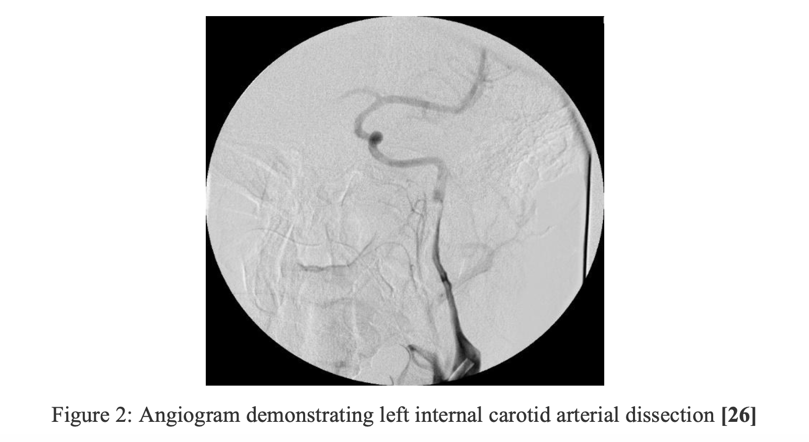
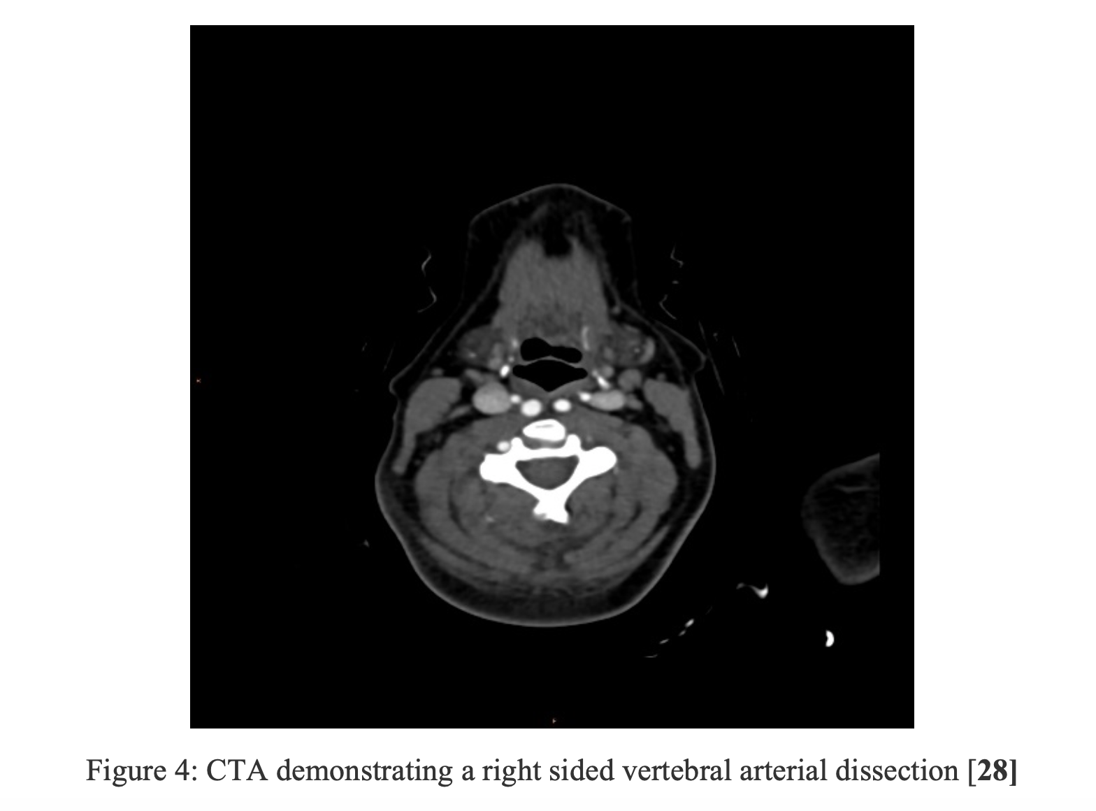
There are several imaging modalities used to diagnose cervical artery dissection. The gold standard for diagnosis of CAD is digital subtraction angiography, but it is rarely used. [19] CTA and MRA are acceptable alternatives to the gold standard. CTA extracranial/ intracranial vessels were shown to be better at visualizing complications of dissection when compared to MRA and were preferred for the diagnosis of vertebral artery dissections [19] The vascular US can be used as an adjunct for initial diagnosis and serial imaging although it has poor sensitivity in detecting cervical artery dissection on its own. [20,21]
Authors of one retrospective study compared test characteristics of CTA and MRA imaging for diagnosis of CAD by comparing calculated test performance characteristics of twenty-one different articles. CTA studies had sensitivities ranging from 51-100% and specificities ranging from 67-100%. [21] Positive predictive values for CTA studies ranged from 65-100% and negative predictive values for CTA ranged from 70-100%. The studies included both prospective and retrospective sub-types and the authors concluded that the ranges were similar between retrospective and prospective CTA studies. [21] In the included MRA studies, the sensitivity values of the studies ranged from 50-100% and the specificity values ranged from 67-99%. The authors found only one paper that calculated PPV and NPV in the MRA literature so they could not compare test performance characteristics between retrospective and prospective studies. [21] In one retrospective study, vascular US was shown to have a sensitivity ranging between 38-86% overall in detecting cervical artery dissections. [22]
In a prospective study with 60 patients, the authors looked at the test characteristics of helical CTA for diagnosis of dissection in patients with penetrating trauma to the neck and found that CTA had a specificity of 100% and a sensitivity of 90% indicating the usefulness of this test for quick and accurate diagnosis in these scenarios when there is concern for dissection. Conventional angiography was used as the gold standard in these patients. [23]
In Vertinsky et al, the authors compared the ability of CTA and MRA imaging modalities in detecting abnormalities commonly seen in cervical artery dissections. [8] Findings that can be detected in CTA and MRA studies include acute ischemic stroke, pseudo-aneurysm, intimal flap, intramural hematoma, vessel stenosis and/or occlusion, and vessel wall abnormalities. In 17 out of the 18 cases in the study, CTA extracranial/intracranial vessels were preferred by the radiologists due to its ability to identify pathologic findings including intimal flaps, pseudo-aneurysms and the presence of a “string sign” (residual patent vessel lumen). [19] CTA studies identified intimal flaps in seven vessels where none were seen on MRA. CTA studies also picked up six pseudo-aneurysms where the MRA studies only picked up two (33%) of these pseudo-aneurysms. In six imaging studies where the MRA read a vessel as occluded, CTA imaging demonstrated a “string sign” in three (50 %) of these cases. In the diagnosis of vertebral artery dissection, CTA was the preferred imaging modality. [19] MRA was superior at detecting acute ischemic stroke when compared to CTA. [19] Both imaging modalities are able to pick up an intramural hematoma in different ways. On CTA, the presence of wall thickening suggests the presence of an intramural hematoma, although it is not very specific when compared to the presence of a methemoglobin crescent seen on MRA (axial T1- weighted fat-suppressed image). [19] Although this study had several limitations (unblinding of neuro-radiologists to clinical presentation and order of imaging studies, lack of control cases, separation of time between some of the imaging studies), this study suggests that in cases where CAD is suspected, CTA might be a better initial modality than MRA to pick up subtle pathologic findings that can guide acute intervention for these patients. CTA also seems to be preferred in cases where vertebral artery dissections are suspected. MRA is superior at detecting signs of acute ischemic stroke in patients with a focal neurological exam. Despite these findings, CTA and MRA can be used together to confirm different findings in patients with complex cases and both can guide potential treatment options. [19]
Another possible option is the vascular US. In a small 1995 prospective cohort study with 44 patients, the author looked at the use of vascular US for work up of cervical artery dissections. [20] The vascular US was not only able to confirm the diagnosis but used to follow the progression of the disease. Doppler and duplex ultrasonography were able to detect the presence of cervical artery dissection in 91.5 % of patients where there was high clinical suspicion for the disease and in 96% of the patients who had evidence of a significant occlusion and/or stenosis in the vessels. Using US for serial studies demonstrated all of the ultrasound studies were verified using MRI/MRA imaging of the vessels. This shows that Doppler and/or duplex studies can potentially serve as an adjunct for initial imaging in patients with a high pre-test probability for cervical artery dissection but it should not be used as a primary imaging modality at this time. The study was small, all of the US findings had to be verified by other imaging modalities and ultrasound is user-dependent so more studies should be done before ultrasound studies can be used alone for diagnosing and following the progression of the disease. [20]
Debate of the Gold standard: Digital subtraction angiography has always been the conventional gold standard for diagnosis of cervical artery dissection but MRA has been replacing this method as a possible new emerging gold standard. [8,19] MR angiography with fat suppression techniques can not only identify the presence of an intramural hematoma but it has been shown to be superior to angiography in identifying dissection in cases with non-specific occlusions and in absence of luminal abnormalities in the affected artery. [10] On the other hand, CTA seems to be superior when it comes to detecting vertebral artery dissection when compared to MRA. [23] In one prospective study, a trauma institution that switched from using conventional angiography to CTA-alone based screening/diagnosis in blunt trauma saw a decrease in time to diagnosis of CAD time from around 31.2 hours to 2.6 hours (p <0.001) and a decrease in overall stroke risk from 15.2% to 3.8%. [24] Yet, at this time, digital subtraction angiography remains the gold standard until more studies are completed comparing the three imaging modalities. [25]

Pearls/Pitfalls:
2) Although conventional angiography is the gold standard, both CTA and MRA can be used for the diagnosis of CAD. Consider using CTA over MRA if concern for vertebral artery dissection.
3) If your patient has concerning findings for CAD with negative CTA and/or MRA results, DO NOT stop your work-up. Proceed to conventional angiography (gold standard).
Treatment:
CAD increases the risk of thromboembolic events. Treatment regimens including both anti-platelet and anti-coagulation drugs have been used to prevent these occurrences. In the 2015 multi-center RCT, CADISS, the authors compared anti-platelet vs anticoagulant drugs for 3 months in patients diagnosed with CAD. [29] The primary endpoints were the development of an ipsilateral stroke or death in the intention-to-treat population. Strokes or death occurred in 2% of the anti-platelet group vs 1% of the anticoagulation group (OR 0.335, p= 0.63) but the results were not statistically significant and overall had a low occurrence rate. They concluded that there was no significant difference in efficacy at preventing stroke or death in patients with symptomatic dissection disease (vertebral and carotid) and overall stroke risk was low. [29]
In a 2009 retrospective study, the authors looked at the outcomes for patients who were given IV thrombolytics caused by CAD vs non- CAD causes. Although the CAD group did not recover as well after 3 months when compared to the non-CAD group, the rates of intracranial hemorrhage and recurrent ischemic groups were equal amongst both groups. [30] In another multi-center retrospective study, the authors compared outcomes in patients with cervical artery dissections complicated by stroke who received thrombolytic therapy and patients who did not receive thrombolytic therapy. The likelihood for favorable outcome based on the modified Rankin scale (0-2) and the occurrence of adverse effects (any intra-cranial hemorrhage and/or any major extracranial hemorrhage) did not differ significantly between the two groups. [31] Both of these papers suggest that thrombolytics should not be withheld in patients with stroke due to CAD who could potentially benefit from this intervention, but the therapy also does not have any clear benefit within this group. Thrombolysis is contraindicated in patients with intracranial extension of the dissection or a dissection involving the aorta due to risk of aortic rupture and/or subarachnoid hemorrhage. [32]
In one small prospective cohort study, the researchers looked at the efficacy of stent angioplasty in symptomatic patients with critical stenosis of vessels due to dissection and found that there are possible benefits for some high-risk patients (failed medical management, critical stenosis, anticoagulation contraindicated). [33] The study only had a sample size of 10 and several of the patients included in the study needed multiple stents so this treatment option still requires further investigation for lower risk CAD patients as a primary treatment intervention. [33] A prospective study done in 2000, evaluated the indications for surgical intervention (vessel reconstruction, clipping) in symptomatic CAD. 48 patients diagnosed with CAD were followed. The surgical intervention occurred for patients with failed medical management on anticoagulation for 6 months, persistent high-grade carotid-stenosis and/or persistent or newly developed carotid aneurysms. Although only one patient died (due to intracranial bleeding), 5 patients (10%) in the study developed minor recurrent stroke and 29 patients (58%) developed cranial nerve damage indicating that surgical intervention is in no way a benign alternative. [34]
The accepted duration of treatment is three to six months although the optimal duration is unclear. Repeat vascular imaging is needed after treatment to look for improvement of cervical artery dissection complications. [8] The vascular US can be used as an adjunct for serial imaging. [20,22]
Pearls/Pitfalls:
4) There is no difference in efficacy between anti-platelet and anticoagulation therapy in patients with symptomatic CAD (CADISS trial).
5) Accepted treatment duration is 3-6 months although the optimal duration is unclear. Repeat vascular imaging is required.
Case Conclusion:
After a history and physical exam are completed, cervical artery dissection is suspected. A CTA of his extracranial and intracranial arteries was ordered which showed a left extracranial internal carotid artery dissection. The patient was admitted to the hospital for further management and observation. The patient had an unremarkable hospital course and was discharged on anti-platelet therapy with outpatient follow up. His symptoms resolved over the next couple of weeks. Repeat vascular imaging several months later showed adequate recanalization of the dissected vessel.
References/Further Reading
1) Figure 1- Case courtesy of Assoc Prof Frank Gaillard, <ahref=”https://radiopaedia.org/”>Radiopaedia.org</a>. From the case <a href=”https://radiopaedia.org/cases/6693″>rID: 6693</a>
2) Schievink W, Mokri B, Whisnant J. Internal carotid artery dissection in a community. Rochester, Minnesota, 1987-1992. Stroke. 1993; 24(11):1678-1680.
3) Lee V, Brown R, Mandrekar J, Mokri B. Incidence and outcome of cervical artery dissection: A population-based study. Neurology. 2006; 67(10):1809-1812.
4) Arnold M, Kappeler L, Georgiadis D et al. Gender differences in spontaneous cervical artery dissection. Neurology. 2006; 67(6):1050-1052.
5) Blum C, Yaghi S. Cervical Artery Dissection: A Review of the Epidemiology, Pathophysiology, Treatment, and Outcome. Arch Neurosci. 2015; 2(4).
6) Morris N, Merkler A, Gialdini G, Kamel H. Timing of Incident Stroke Risk After Cervical Artery Dissection Presenting Without Ischemia. Stroke. 2017; 48(3):551-555.
7) Rubinstein S, Peerdeman S, van Tulder M, Riphagen I, Haldeman S. A Systematic Review of the Risk Factors for Cervical Artery Dissection. Stroke. 2005; 36(7):1575-1580.
8) Thanvi B. Carotid and vertebral artery dissection syndromes. Postgrad Med J. 2005; 81(956):383-388.
9) Brandt T, Orbek E, Hacke W. Cervical artery dissection syndromes. In: Bogousslavsky J, Caplan L, eds. Stroke syndromes. 2nd ed. Cambridge: Cambridge University Press, 2001:660–6.
10) Schievink W. I. Spontaneous dissection of the carotid and vertebral arteries. N Engl J Med 2001; 344:898–906.
11) Benninger DH, Georgiadia D, Kremer C. Mechanism of ischaemic infarction in spontaneous carotid dissection. Stroke 2004; 35:482–5. 29
12) Baumgartner RW, Bogousslavsky J. Clinical manifestations of carotid dissection. Front Neurol Neurosci. 2005; 20:70-76.
13) Biousse V, Schaison M, Touboul P-J, et al. Ischaemic optic neuropathy associated with internal carotid artery dissection. Arch Neurol 1998; 55:715–19.
14) Tabatabai G, Schöber W, Ernemann U, et al. Vertebral artery dissection presenting with ipsilateral acute C5 and C6 sensorimotor radiculopathy: A case report. Cases J. 2008; 1:139-139.
15) Arnold M, Bousser M, Fahrni G et al. Vertebral Artery Dissection. Stroke. 2006; 37(10):2499-2503.
16) Debette S, Grond-Ginsbach C, Bodenant M et al. Differential features of carotid and vertebral artery dissections: The CADISP Study. Neurology. 2011; 77(12):1174-1181.
17) Arnold M. Pain as the only symptom of cervical artery dissection. Journal of Neurology, Neurosurgery & Psychiatry. 2006; 77(9):1021-1024.
18) Hardmeier M, Haller S, Steck A, et al. Vertebral artery dissection presenting with fifth cervical root (C5) radiculopathy. J Neurol. 2007; 254(5):672-673.
19) Vertinsky A, Schwartz N, Fischbein N, Rosenberg J, Albers G, Zaharchuk G. Comparison of Multidetector CT Angiography and MR Imaging of Cervical Artery Dissection. American Journal of Neuroradiology. 2008; 29(9):1753-1760.
20) Sturzenegger M. Spontaneous internal carotid artery dissection: Early diagnosis and management in 44 patients. J Neurol. 1995; 242(4):231-238.
21) Provenzale J, Sarikaya B. Comparison of Test Performance Characteristics of MRI, MR Angiography, and CT Angiography in the Diagnosis of Carotid and Vertebral Artery Dissection: A Review of the Medical Literature. American Journal of Roentgenology. 2009; 193: 1167-1174.
22) Mutze S, Rademacher G, Matthes G, et al. Blunt cerebrovascular injury in patients with blunt multiple trauma: diagnostic accuracy of duplex Doppler US and early CT angiography. Radiology. 2005; 237(3):884-892.
23) Munera F, Soto JA, Palacio D, et al. Diagnosis of arterial injuries caused by penetrating trauma to the neck: comparison of helical CT angiography and conventional angiography. Radiology. 2000; 216(2):356-362.
24) Eastman AL, Muraliraj V, Sperry JL, et al. CTA-based screening reduces time to diagnosis and stroke rate in blunt cervical vascular injury. J Trauma. 2009; 67(3):551-556.
25) Rodallec M, Marteau V, Gerber S, Desmottes L, Zins M. Craniocervical Arterial Dissection: Spectrum of Imaging Findings and Differential Diagnosis. RadioGraphics. 2008; 28 (6).
26) Figure 2 – Case courtesy of Assoc Prof Frank Gaillard, <ahref=”https://radiopaedia.org/”>Radiopaedia.org</a>. From the case <a href=”https://radiopaedia.org/cases/5182″>rID: 5182</a>
27) Figure 3- MRA showing right carotid arterial dissection, Case courtesy of Dr Mohammad A. ElBeialy, <a href=”https://radiopaedia.org/”>Radiopaedia.org</a>. From the case <a href=”https://radiopaedia.org/cases/26636″>rID: 26636</a>
28) Figure 4: CTA showing vertebral artery dissection, Case courtesy of Dr Derek Smith, <a href=”https://radiopaedia.org/”>Radiopaedia.org</a>. From the case <a href=”https://radiopaedia.org/cases/57166″>rID: 57166</a>
29) Markus H, Hayter E, Levi C, Feldman A, Venables G, Norris J. Antiplatelet treatment compared with anticoagulation treatment for cervical artery dissection (CADISS): a randomised trial. The Lancet Neurology. 2015; 14(4):361-367.
30) Engelter S, Rutgers M, Hatz F et al. Intravenous Thrombolysis in Stroke Attributable to Cervical Artery Dissection. Stroke. 2009; 40(12):3772-3776.
31) Engelter S, Dallongeville J, Kloss M et al. Thrombolysis in Cervical Artery Dissection – Data from the Cervical Artery Dissection and Ischaemic Stroke Patients (CADISP) database. Eur J Neurol. 2012; 19(9):1199-1206.
32) Patel R, Adam R, Malkjian C, et al. Cervical carotid artery dissection: current review of diagnosis and treatment. Cardiology in Review. 2012; 20(3): 145-152.
33) Malek A, Higashida R, Phatouros C et al. Stent Angioplasty for Cervical Carotid Artery Stenosis in High-Risk Symptomatic NASCET-Ineligible Patients. Stroke. 2000; 31(12):3029-3033.
34) Müller B, Luther B, Hort W, Neumann-Haefelin T, Aulich A, Sandmann W. Surgical treatment of 50 carotid dissections: Indications and results. J Vasc Surg. 2000; 31(5):980-988.
35) Levac-Martinho O, Simon E. EM@3AM – Carotid Artery Dissection – emDOCs.net – Emergency Medicine Education. emDOCs.net – Emergency Medicine Education. http://www.emdocs.net/em3am-carotid-artery-dissection/. Published 2020. Accessed February 5, 2020.
36) Schievink W. I. Spontaneous Dissection of the Carotid and Vertebral Arteries. New England Journal of Medicine. 2001; 344:898-906.
37) Peng J, Liu Z, Luo C et al. Treatment of Cervical Artery Dissection: Antithrombotics, Thrombolysis, and Endovascular Therapy. Biomed Res Int. 2017; 2017:1-6.
38) Akala O. Carotid Artery Dissection – emDOCs.net – Emergency Medicine Education. emDOCs.net – Emergency Medicine Education. http://www.emdocs.net/carotid-artery-dissection/. Published 2020. Accessed February 5, 2020.
39) Cadena R. Cervical Artery Dissection in the Emergency Room: Symptoms, Diagnosis, Treatment. Ebmedicine.net. https://www.ebmedicine.net/topics/cardiovascular/cervical-artery-dissection. Published 2020. Accessed February 19, 2020.


